Explore
 Validate
 Learn
Learn Western blot
Antibody data
- Antibody Data
- Antigen structure
- References [2]
- Comments [0]
- Validations
- Western blot [3]
- Immunohistochemistry [1]
Submit
Validation data
Reference
Comment
Report error
- Product number
- AF4094 - Provider product page

- Provider
- R&D Systems
- Product name
- Human/Mouse/Rat FKBP51 Antibody
- Antibody type
- Polyclonal
- Description
- Antigen Affinity-purified. Detects human, mouse, and rat FKBP51. In Western blots, less than 1% cross-reactivity with recombinant human FKBP12, FKBP13, FKBP38, and FKBP52 is observed.
- Reactivity
- Human, Mouse, Rat
- Host
- Goat
- Conjugate
- Unconjugated
- Antigen sequence
Q13451- Isotype
- IgG
- Vial size
- 100 ug
- Concentration
- LYOPH
- Storage
- Use a manual defrost freezer and avoid repeated freeze-thaw cycles. 12 months from date of receipt, -20 to -70 °C as supplied. 1 month, 2 to 8 °C under sterile conditions after reconstitution. 6 months, -20 to -70 °C under sterile conditions after reconstitution.
Submitted references Unique Gene Expression Signatures in the Intestinal Mucosa and Organoids Derived from Germ-Free and Monoassociated Mice.
Glucocorticoid-regulated genes in eosinophilic esophagitis: a role for FKBP51.
Janeckova L, Kostovcikova K, Svec J, Stastna M, Strnad H, Kolar M, Hudcovic T, Stancikova J, Tureckova J, Baloghova N, Sloncova E, Galuskova K, Tlaskalova-Hogenova H, Korinek V
International journal of molecular sciences 2019 Mar 29;20(7)
International journal of molecular sciences 2019 Mar 29;20(7)
Glucocorticoid-regulated genes in eosinophilic esophagitis: a role for FKBP51.
Caldwell JM, Blanchard C, Collins MH, Putnam PE, Kaul A, Aceves SS, Bouska CA, Rothenberg ME
The Journal of allergy and clinical immunology 2010 Apr;125(4):879-888.e8
The Journal of allergy and clinical immunology 2010 Apr;125(4):879-888.e8
No comments: Submit comment
Supportive validation
- Submitted by
- R&D Systems (provider)
- Main image
- Experimental details
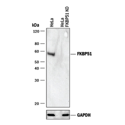
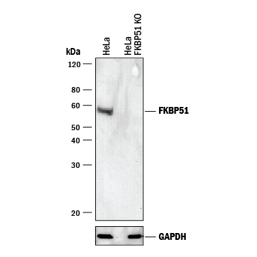
- Western Blot Shows Human FKBP51 Specificity by Using Knockout Cell Line. Western blot shows lysates of HeLa human cervical epithelial carcinoma parental cell line and FKBP51 knockout HeLa cell line (KO). PVDF membrane was probed with 0.3 µg/mL of Goat Anti-Human/Mouse/Rat FKBP51 Antigen Affinity-purified Polyclonal Antibody (Catalog # AF4094) followed by HRP-conjugated Anti-Mouse IgG Secondary Antibody (Catalog # HAF018). A specific band was detected for FKBP51 at approximately 32 kDa (as indicated) in the parental HeLa cell line, but is not detectable in knockout HeLa cell line. GAPDH (Catalog # AF5718) is shown as a loading control. This experiment was conducted under reducing conditions and using Immunoblot Buffer Group 1.
- Submitted by
- R&D Systems (provider)
- Main image

- Experimental details
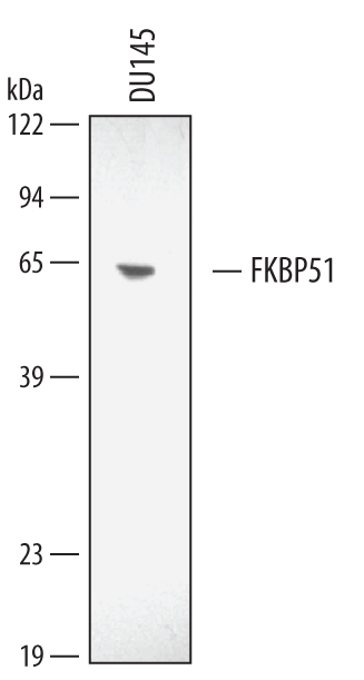
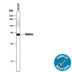
- Detection of Human/Mouse/Rat FKBP51 by Western Blot. Western blot shows lysates of DU145 human prostate carcinoma cell line. PVDF membrane was probed with 0.3 µg/mL of Human/Mouse/Rat FKBP51 Antigen Affinity-purified Polyclonal Antibody (Catalog # AF4094) followed by HRP-conjugated Anti-Goat IgG Secondary Antibody (Catalog # HAF109). A specific band was detected for FKBP51 at approximately 51 kDa (as indicated). This experiment was conducted under reducing conditions and using Immunoblot Buffer Group 2.
- Submitted by
- R&D Systems (provider)
- Main image
- Experimental details
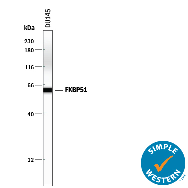
- Detection of Human FKBP51 by Simple WesternTM. Simple Western lane view shows lysates of DU145 human prostate carcinoma cell line, loaded at 0.2 mg/mL. A specific band was detected for FKBP51 at approximately 61 kDa (as indicated) using 3 µg/mL of Goat Anti-Human/Mouse/Rat FKBP51 Antigen Affinity-purified Polyclonal Antibody (Catalog # AF4094) followed by 1:50 dilution of HRP-conjugated Anti-Goat IgG Secondary Antibody (Catalog # HAF109). This experiment was conducted under reducing conditions and using the 12-230 kDa separation system.
Supportive validation
- Submitted by
- R&D Systems (provider)
- Main image
- Experimental details
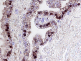
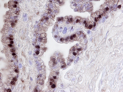
- FKBP51 in Human Prostate. FKBP51 was detected in immersion fixed paraffin-embedded sections of human prostate using 15 µg/mL Human/Mouse/Rat FKBP51 Antigen Affinity-purified Polyclonal Antibody (Catalog # AF4094) overnight at 4 °C. Tissue was stained with the Anti-Goat HRP-DAB Cell & Tissue Staining Kit (brown; Catalog # CTS008) and counterstained with hematoxylin (blue). View our protocol for Chromogenic IHC Staining of Paraffin-embedded Tissue Sections.