Explore
 Validate
 Learn
Learn Western blot
 Other assay
Antibody data
- Antibody Data
- Antigen structure
- References [1]
- Comments [0]
- Validations
- Other assay [1]
Submit
Validation data
Reference
Comment
Report error
- Product number
- BMS1044FI - Provider product page

- Provider
- Invitrogen Antibodies
- Product name
- IFN beta Monoclonal Antibody (A1 (IFNb)), FITC, eBioscience™
- Antibody type
- Monoclonal
- Antigen
- Other
- Description
- Description: The IFNb/A1 clone recognizes natural human IFN-beta and recombinants beta1a and beta1b. It is capable of neutralizing IFN-beta. Interferon beta is synthesized and secreted by fibroblasts and many other cell types in response to pathogens. IFN-beta binding to type I interferon receptors induces the upregulation of IRF-7 and activation of Rnase L. IRF-7 can exert a positive feedback on IFN-beta production. RNase L cleaves both viral and cellular single stranded mRNA, thereby limiting viral replication and dissemination. Applications Tested: ELISA, Neutralization tests, Western Blotting. Excitation: 488 nm; Emission: 520 nm; Laser: Blue Laser
- Reactivity
- Human
- Host
- Mouse
- Conjugate
- Green dye
- Isotype
- IgG
- Antibody clone number
- A1 (IFNb)
- Vial size
- 100 µg
- Concentration
- 0.1 mg/mL
- Storage
- 4° C, store in dark
Submitted references Deubiquitination of proteasome subunits by OTULIN regulates type I IFN production.
Tao P, Wang S, Ozen S, Lee PY, Zhang J, Wang J, Han H, Yang Z, Fang R, Tsai WL, Yang H, Sag E, Topaloglu R, Aksentijevich I, Yu X, Zhou Q
Science advances 2021 Nov 19;7(47):eabi6794
Science advances 2021 Nov 19;7(47):eabi6794
No comments: Submit comment
Supportive validation
- Submitted by
- Invitrogen Antibodies (provider)
- Main image

- Experimental details
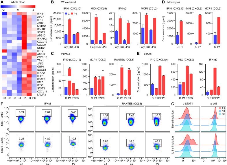
- Fig. 1. Patients with otulipenia display overactivation of IFN-I signaling. ( A ) NanoString analysis of IFN-I signaling in whole blood samples from two patients, four healthy controls, and a type I interferonopathy patient control (PC) with deoxyribonuclease 2 deficiency. ( B ) Cytokine levels in whole blood samples. The whole blood samples from P1 and his unaffected sibling were stimulated with poly(I:C) (20 mug/ml) or LPS (1 mug/ml) for 22 hours. ( C ) Cytokine levels in the supernatant of cultured PBMCs from three patients and one healthy control. ( D ) Cytokine levels in the supernatant of purified monocytes from P1 and three healthy controls. Cells used to detect IP10 and monokine induced by IFN-gamma (MIG) were at basal level. Cells used to detect monocyte chemoattractant protein 1 (MCP1) were stimulated with LPS (1 mug/ml) for 48 hours. P1 was sampled twice. ( E ) Serum cytokine levels from three patients and seven healthy controls. ( F ) Intracellular cytokine staining of IFN-beta and RANTES (CCL5) in T cells and B cells of P3 and two healthy controls. ( G ) Phosphorylation of STAT1 and p65 of patient P3 T cells compared to two healthy controls at basal level and after IL-6 (100 ng/ml) stimulation for 6 hours as determined by flow cytometry analysis. The patients carry homozygous disease-associated variants in OTULIN: P1 (Leu 272 Pro), P2 (Tyr 244 Cys), and P3 (Gly 174 Aspfs*2).
- Conjugate
- Green dye