Explore
 Validate
 Learn
Learn Western blot
 Immunohistochemistry
Antibody data
- Antibody Data
- Antigen structure
- References [19]
- Comments [0]
- Validations
- Immunohistochemistry [1]
- Other assay [5]
Submit
Validation data
Reference
Comment
Report error
- Product number
- 32-6000 - Provider product page

- Provider
- Invitrogen Antibodies
- Product name
- Desmoglein 1 Monoclonal Antibody (27B2)
- Antibody type
- Monoclonal
- Antigen
- Other
- Reactivity
- Human
- Host
- Mouse
- Isotype
- IgG
- Antibody clone number
- 27B2
- Vial size
- 100 μg
- Concentration
- 0.5 mg/mL
- Storage
- -20°C
Submitted references FAM167A is a key molecule to induce BCR-ABL-independent TKI resistance in CML via noncanonical NF-κB signaling activation.
Elemental and molecular imaging of human full thickness skin after exposure to heavy metals.
In vitro Characteristics of Heterogeneous Equine Hoof Progenitor Cell Isolates.
Fisetin, a 3,7,3',4'-Tetrahydroxyflavone Inhibits the PI3K/Akt/mTOR and MAPK Pathways and Ameliorates Psoriasis Pathology in 2D and 3D Organotypic Human Inflammatory Skin Models.
The Differentiation-Associated Keratinocyte Protein Cornifelin Contributes to Cell-Cell Adhesion of Epidermal and Mucosal Keratinocytes.
The cell-cell junctions of mammalian testes: II. The lamellar smooth muscle monolayer cells of the peritubular wall are laterally connected by vertical adherens junctions-a novel architectonic cell-cell junction system.
Role of Fibroblast Growth Factor Receptor 2b in the Cross Talk between Autophagy and Differentiation: Involvement of Jun N-Terminal Protein Kinase Signaling.
A non-canonical role for desmoglein-2 in endothelial cells: implications for neoangiogenesis.
Early dental epithelial transcription factors distinguish ameloblastoma from keratocystic odontogenic tumor.
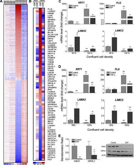
Alteration of the EphA2/Ephrin-A signaling axis in psoriatic epidermis.
Desmoglein-1/Erbin interaction suppresses ERK activation to support epidermal differentiation.
Assessment of desmosomal components (desmoglein 1-3, plakoglobin) in cardia mucosa in relation to gastroesophageal reflux disease and Helicobacter pylori infection.
Gastro-oesophageal reflux disease is associated with up-regulation of desmosomal components in oesophageal mucosa.
Hailey-Hailey disease and tight junctions: Claudins 1 and 4 are regulated by ATP2C1 gene encoding Ca(2+) /Mn(2+) ATPase SPCA1 in cultured keratinocytes.
EGFR regulation of epidermal barrier function.
Ligand targeting of EphA2 enhances keratinocyte adhesion and differentiation via desmoglein 1.
Human eccrine sweat gland cells can reconstitute a stratified epidermis.
P120 catenin is associated with desmogleins when desmosomes are assembled in high-Ca2+ medium but not when disassembled in low-Ca2+ medium in DJM-1 cells.
New skin-equivalent model from de-epithelialized amnion membrane.
Yang T, Sim KY, Ko GH, Ahn JS, Kim HJ, Park SG
Journal of experimental & clinical cancer research : CR 2022 Mar 3;41(1):82
Journal of experimental & clinical cancer research : CR 2022 Mar 3;41(1):82
Elemental and molecular imaging of human full thickness skin after exposure to heavy metals.
Chavatte L, Juan M, Mounicou S, Leblanc Noblesse E, Pays K, Nizard C, Bulteau AL
Metallomics : integrated biometal science 2020 Oct 21;12(10):1555-1562
Metallomics : integrated biometal science 2020 Oct 21;12(10):1555-1562
In vitro Characteristics of Heterogeneous Equine Hoof Progenitor Cell Isolates.
Yang Q, Pinto VMR, Duan W, Paxton EE, Dessauer JH, Ryan W, Lopez MJ
Frontiers in bioengineering and biotechnology 2019;7:155
Frontiers in bioengineering and biotechnology 2019;7:155
Fisetin, a 3,7,3',4'-Tetrahydroxyflavone Inhibits the PI3K/Akt/mTOR and MAPK Pathways and Ameliorates Psoriasis Pathology in 2D and 3D Organotypic Human Inflammatory Skin Models.
Chamcheu JC, Esnault S, Adhami VM, Noll AL, Banang-Mbeumi S, Roy T, Singh SS, Huang S, Kousoulas KG, Mukhtar H
Cells 2019 Sep 15;8(9)
Cells 2019 Sep 15;8(9)
The Differentiation-Associated Keratinocyte Protein Cornifelin Contributes to Cell-Cell Adhesion of Epidermal and Mucosal Keratinocytes.
Wagner T, Beer L, Gschwandtner M, Eckhart L, Kalinina P, Laggner M, Ellinger A, Gruber R, Kuchler U, Golabi B, Tschachler E, Mildner M
The Journal of investigative dermatology 2019 Nov;139(11):2292-2301.e9
The Journal of investigative dermatology 2019 Nov;139(11):2292-2301.e9
The cell-cell junctions of mammalian testes: II. The lamellar smooth muscle monolayer cells of the peritubular wall are laterally connected by vertical adherens junctions-a novel architectonic cell-cell junction system.
Domke LM, Franke WW
Cell and tissue research 2019 Feb;375(2):451-482
Cell and tissue research 2019 Feb;375(2):451-482
Role of Fibroblast Growth Factor Receptor 2b in the Cross Talk between Autophagy and Differentiation: Involvement of Jun N-Terminal Protein Kinase Signaling.
Nanni M, Ranieri D, Rosato B, Torrisi MR, Belleudi F
Molecular and cellular biology 2018 Jul 1;38(13)
Molecular and cellular biology 2018 Jul 1;38(13)
A non-canonical role for desmoglein-2 in endothelial cells: implications for neoangiogenesis.
Ebert LM, Tan LY, Johan MZ, Min KK, Cockshell MP, Parham KA, Betterman KL, Szeto P, Boyle S, Silva L, Peng A, Zhang Y, Ruszkiewicz A, Zannettino AC, Gronthos S, Koblar S, Harvey NL, Lopez AF, Shackleton M, Bonder CS
Angiogenesis 2016 Oct;19(4):463-86
Angiogenesis 2016 Oct;19(4):463-86
Early dental epithelial transcription factors distinguish ameloblastoma from keratocystic odontogenic tumor.
Heikinheimo K, Kurppa KJ, Laiho A, Peltonen S, Berdal A, Bouattour A, Ruhin B, Catón J, Thesleff I, Leivo I, Morgan PR
Journal of dental research 2015 Jan;94(1):101-11
Journal of dental research 2015 Jan;94(1):101-11
Alteration of the EphA2/Ephrin-A signaling axis in psoriatic epidermis.
Gordon K, Kochkodan JJ, Blatt H, Lin SY, Kaplan N, Johnston A, Swindell WR, Hoover P, Schlosser BJ, Elder JT, Gudjonsson JE, Getsios S
The Journal of investigative dermatology 2013 Mar;133(3):712-722
The Journal of investigative dermatology 2013 Mar;133(3):712-722
Desmoglein-1/Erbin interaction suppresses ERK activation to support epidermal differentiation.
Harmon RM, Simpson CL, Johnson JL, Koetsier JL, Dubash AD, Najor NA, Sarig O, Sprecher E, Green KJ
The Journal of clinical investigation 2013 Apr;123(4):1556-70
The Journal of clinical investigation 2013 Apr;123(4):1556-70
Assessment of desmosomal components (desmoglein 1-3, plakoglobin) in cardia mucosa in relation to gastroesophageal reflux disease and Helicobacter pylori infection.
Wex T, Kuester D, Mönkemüller K, Stahr A, Fry LC, Kandulski A, Kropf S, Roessner A, Malfertheiner P
Human pathology 2012 Oct;43(10):1745-54
Human pathology 2012 Oct;43(10):1745-54
Gastro-oesophageal reflux disease is associated with up-regulation of desmosomal components in oesophageal mucosa.
Wex T, Mönkemüller K, Stahr A, Kuester D, Fry LC, Völkel S, Kandulski A, Roessner A, Malfertheiner P
Histopathology 2012 Feb;60(3):405-15
Histopathology 2012 Feb;60(3):405-15
Hailey-Hailey disease and tight junctions: Claudins 1 and 4 are regulated by ATP2C1 gene encoding Ca(2+) /Mn(2+) ATPase SPCA1 in cultured keratinocytes.
Raiko L, Siljamäki E, Mahoney MG, Putaala H, Suominen E, Peltonen J, Peltonen S
Experimental dermatology 2012 Aug;21(8):586-91
Experimental dermatology 2012 Aug;21(8):586-91
EGFR regulation of epidermal barrier function.
Tran QT, Kennedy LH, Leon Carrion S, Bodreddigari S, Goodwin SB, Sutter CH, Sutter TR
Physiological genomics 2012 Apr 15;44(8):455-69
Physiological genomics 2012 Apr 15;44(8):455-69
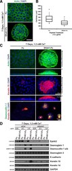
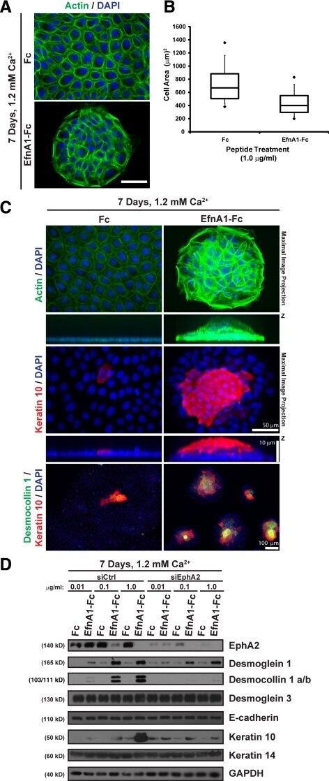
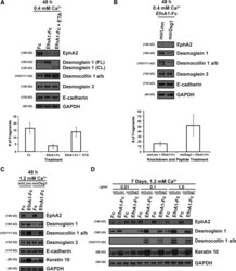
Ligand targeting of EphA2 enhances keratinocyte adhesion and differentiation via desmoglein 1.
Lin S, Gordon K, Kaplan N, Getsios S
Molecular biology of the cell 2010 Nov 15;21(22):3902-14
Molecular biology of the cell 2010 Nov 15;21(22):3902-14
Human eccrine sweat gland cells can reconstitute a stratified epidermis.
Biedermann T, Pontiggia L, Böttcher-Haberzeth S, Tharakan S, Braziulis E, Schiestl C, Meuli M, Reichmann E
The Journal of investigative dermatology 2010 Aug;130(8):1996-2009
The Journal of investigative dermatology 2010 Aug;130(8):1996-2009
P120 catenin is associated with desmogleins when desmosomes are assembled in high-Ca2+ medium but not when disassembled in low-Ca2+ medium in DJM-1 cells.
Kanno M, Aoyama Y, Isa Y, Yamamoto Y, Kitajima Y
The Journal of dermatology 2008 Jun;35(6):317-24
The Journal of dermatology 2008 Jun;35(6):317-24
New skin-equivalent model from de-epithelialized amnion membrane.
Yang L, Shirakata Y, Shudou M, Dai X, Tokumaru S, Hirakawa S, Sayama K, Hamuro J, Hashimoto K
Cell and tissue research 2006 Oct;326(1):69-77
Cell and tissue research 2006 Oct;326(1):69-77
No comments: Submit comment
Supportive validation
- Submitted by
- Invitrogen Antibodies (provider)
- Main image
- Experimental details
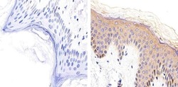
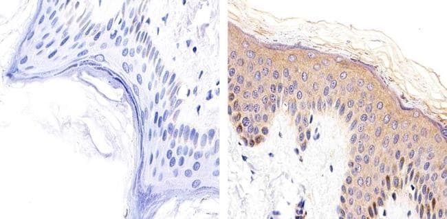
- Immunohistochemistry analysis of Desmoglein-1 showing staining in the cytoplasm and membrane of paraffin-embedded human skin tissue (right) compared to a negative control without primary antibody (left). To expose target proteins, antigen retrieval was performed using 10mM sodium citrate (pH 6.0), microwaved for 8-15 min. Following antigen retrieval, tissues were blocked in 3% H2O2-methanol for 15 min at room temperature, washed with ddH2O and PBS, and then probed with a Desmoglein-1 monoclonal antibody (Product # 32-6000) diluted in 3% BSA-PBS at a dilution of 1:20 overnight at 4ºC in a humidified chamber. Tissues were washed extensively in PBST and detection was performed using an HRP-conjugated secondary antibody followed by colorimetric detection using a DAB kit. Tissues were counterstained with hematoxylin and dehydrated with ethanol and xylene to prep for mounting.
Supportive validation
- Submitted by
- Invitrogen Antibodies (provider)
- Main image
- Experimental details
- NULL
- Submitted by
- Invitrogen Antibodies (provider)
- Main image
- Experimental details
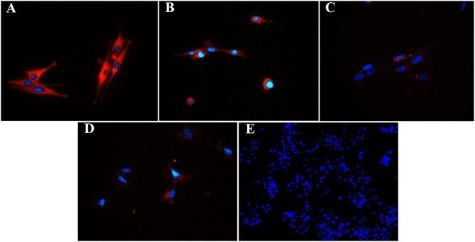
- Figure 3. Ligand targeting of EphA2 triggers keratinocyte colony compaction, stratification and differentiation. (A) Phalloidin (F-actin; in green) and DAPI (nuclei; in blue) staining of sparse keratinocytes that were treated with 1.0 mug/ml Fc or ephrin-A1-Fc for 7 d in 1.2 mM calcium (scale bar, 50 mum). (B) Cell compaction was analyzed by using ImageJ software to measure the surface area of individual cells and presented in a box-and-whisker plot. (C) Maximal image projections (top panels) from peptide-treated colonies that were stained with phalloidin to image F-actin (in green), an antibody against keratin 10 (in red) or DAPI to visualize nuclei (in blue). Apotome-processed images were acquired from stratified keratinocyte colonies at 0.25 mum increments (scale bar, 50 mum). Images below depict cross-sections through the z-x axis that were magnified in the z plane to better resolve keratinocyte piles (scale bar, 10 mum). Colonies that had formed in the presence of Fc or ephrin-A1-Fc peptide in 1.2 mM calcium were also fixed and immunostained for desmocollin 1 a/b (in green) or keratin 10 (in red) to detect stratified keratinocytes (scale bar, 100 mum). (D) siCtrl or siEphA2 keratinocytes were seeded under sparse conditions and treated with 0.01, 0.1, or 1.0 mug/ml Fc or ephrin-A1-Fc peptide for 7 days in 1.2 mM calcium. Western blot analysis of lysates prepared from these cultures were probed for EphA2, desmoglein 1, desmocollin 1 a/b, desmoglein 3, E-cadherin, keratin 1
- Submitted by
- Invitrogen Antibodies (provider)
- Main image
- Experimental details
- Figure 5. Ligand targeting of EphA2 enhances keratinocyte adhesion and differentiation in a desmoglein-1 dependent manner. (A) Confluent keratinocyte sheets were treated with 1.0 mug/ml Fc or ephrin-A1-Fc peptide for 48 h in 0.4 mM calcium and incubated in the presence of 2.0 mug/ml recombinant Staphylococcus aureus exfoliative toxin A (ETA) for an additional 2 h. Top panels show Western blot analysis of EphA2, full length (FL) and cleaved (CL) desmoglein 1, desmocollin 1 a/b, desmoglein 3, E-cadherin, or GAPDH. A mechanical dissociation assay was performed, and the bottom panel contains a bar graph depicting the average number of fragments. (B) Keratinocytes transduced with retroviruses harboring microRNA mimetic sequences targeting lamin A/C (mirLmn) as a control or desmoglein 1 (mirDsg1) were seeded to confluency and treated with 1.0 mug/ml ephrin-A1-Fc peptide for 48 h in 0.4 mM calcium. Top panels show Western blot analysis of EphA2, desmoglein 1, desmocollin 1 a/b, desmoglein 3, E-cadherin, or GAPDH. In parallel, a mechanical dissociation assay was performed (bottom panel). (C) Western blot analysis for EphA2, desmoglein 1, desmocollin 1 a/b, desmoglein 3, E-cadherin, keratin 10, or GAPDH from confluent keratinocytes transduced with mirLmn and mirDsg1 that were treated with 1.0 mug/ml Fc or ephrin-A1-Fc peptide for 48 h in 1.2 mM calcium. (D) Keratinocytes transduced with mirLmn and mirDsg1 were seeded under sparse conditions and treated with 0.01, 0.1, or 1.0 mug/ml Fc

- Submitted by
- Invitrogen Antibodies (provider)
- Main image
- Experimental details
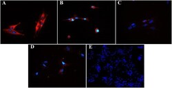
- Figure 8 Fluorescent photomicrographs of cultured progenitor cells (P3) from unaffected (A,B) and laminitic (C,D) hooves labeled (red) with anti- DSG1 (A,C) , DSG3 (B,D) antibodies or no antibodies (E) . DAPI nuclear stain (blue); Magnification = 40X (A-D) ; 20X (E) .
- Submitted by
- Invitrogen Antibodies (provider)
- Main image
- Experimental details
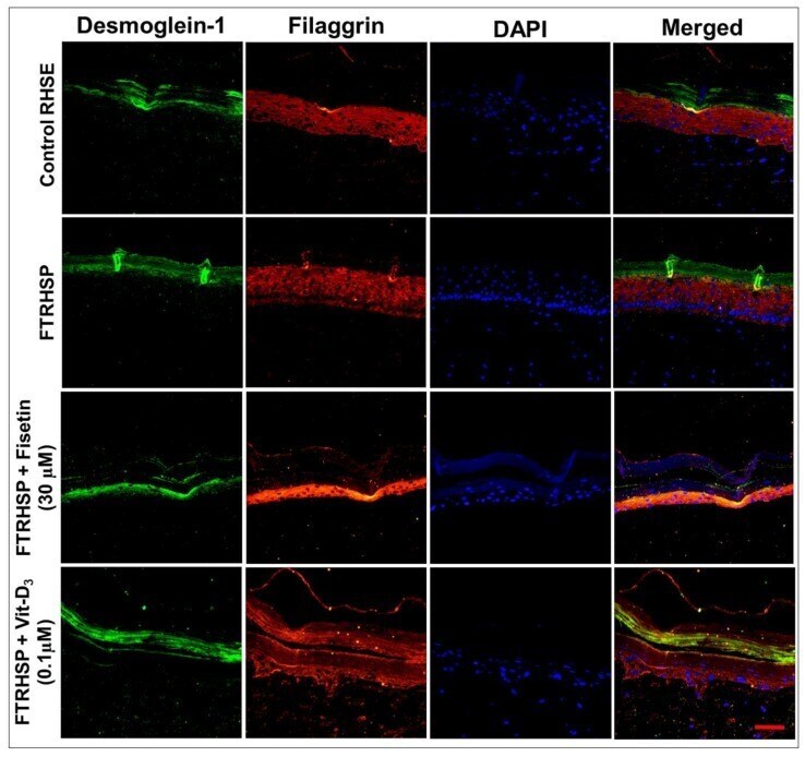
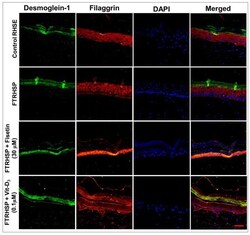
- Figure 9 Representative immunofluorescent photomicrographs showing the protein expression levels of differentiation (filaggrin) and desmosomal (desmoglein-1) protein markers in control RHSE and FTRHSP under different treatment conditions versus fisetin or Vit-D 3 -treated FTRHSP tissues. Results are representative of three independent experiments each performed in quadruplicate and comparing control RHSE vs. FTRHSP treated groups. Data green (Dsgl-1); red (filaggrin), blue (DAPI) and mixed is merged representation. Dsgl-1 = desmoglein-1. Scale bar = 20 mum.