 Explore
 Validate
 Learn
Learn Flow cytometry
Antibody data
- Antibody Data
- Antigen structure
- References [1]
- Comments [0]
- Validations
- Flow cytometry [1]
Submit
Validation data
Reference
Comment
Report error
- Product number
- 12-1429-42 - Provider product page

- Provider
- Invitrogen Antibodies
- Product name
- CD142 Monoclonal Antibody (HTF-1), PE, eBioscience™
- Antibody type
- Monoclonal
- Antigen
- Other
- Description
- Description: This HTF-1 monoclonal antibody reacts with human CD142, which is also known as Tissue Factor. Expression of this type I transmembrane glycoprotein on endothelial cells, monocytes, macrophages, and platelets can be induced by inflammatory mediators (e.g., LPS, IL-1b, TNFa, PMA, or endotoxin). On the other hand, CD142 is expressed constitutively by some tumor cells (e.g., lung, pancreatic, breast, and colon) and non-immune tissues such as the vasculature, central nervous system, kidney, epithelia, and placenta. Studies have also suggested that CD142 exists as a soluble form that circulates in blood. CD142 initiates blood coagulation by associating with and activating the circulating factors VII and VIIa. The HTF-1 antibody has been reported to exhibit blocking activity. Applications Reported: This HTF-1 antibody has been reported for use in flow cytometric analysis. Applications Tested: This HTF-1 antibody has been pre-titrated and tested by flow cytometric analysis of stimulated normal human peripheral blood cells. This can be used at 5 µL (0.125 µg) per test. A test is defined as the amount (µg) of antibody that will stain a cell sample in a final volume of 100 µL. Cell number should be determined empirically but can range from 10^5 to 10^8 cells/test. Excitation: 488-561 nm; Emission: 578 nm; Laser: Blue Laser, Green Laser, Yellow-Green Laser. Filtration: 0.2 µm post-manufacturing filtered.
- Reactivity
- Human
- Host
- Mouse
- Conjugate
- Yellow dye
- Isotype
- IgG
- Antibody clone number
- HTF-1
- Vial size
- 100 Tests
- Concentration
- 5 µL/Test
- Storage
- 4° C, store in dark, DO NOT FREEZE!
Submitted references Tissue factor expression by myeloid cells contributes to protective immune response against Mycobacterium tuberculosis infection.
Venkatasubramanian S, Tripathi D, Tucker T, Paidipally P, Cheekatla S, Welch E, Raghunath A, Jeffers A, Tvinnereim AR, Schechter ME, Andrade BB, Mackman N, Idell S, Vankayalapati R
European journal of immunology 2016 Feb;46(2):464-79
European journal of immunology 2016 Feb;46(2):464-79
No comments: Submit comment
Supportive validation
- Submitted by
- Invitrogen Antibodies (provider)
- Main image
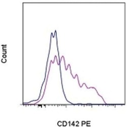
- Experimental details
- Normal human peripheral blood cells were either left unstimulated (blue histogram) or stimulated for 4 hours with LPS (purple histogram) and then stained with Anti-Human CD142 PE. Total viable cells were used for analysis.
- Conjugate
- Yellow dye
