 Explore
 Validate
 Learn
Learn Flow cytometry
Antibody data
- Antibody Data
- Antigen structure
- References [10]
- Comments [0]
- Validations
- Flow cytometry [1]
Submit
Validation data
Reference
Comment
Report error
- Product number
- 17-1429-42 - Provider product page

- Provider
- Invitrogen Antibodies
- Product name
- CD142 Monoclonal Antibody (HTF-1), APC, eBioscience™
- Antibody type
- Monoclonal
- Antigen
- Other
- Description
- Description: This HTF-1 monoclonal antibody reacts with human CD142, which is also known as Tissue Factor. Expression of this type I transmembrane glycoprotein on endothelial cells, monocytes, macrophages, and platelets can be induced by inflammatory mediators (e.g., LPS, IL-1b, TNFa, PMA, or endotoxin). On the other hand, CD142 is expressed constitutively by some tumor cells (e.g., lung, pancreatic, breast, and colon) and non-immune tissues such as the vasculature, central nervous system, kidney, epithelia, and placenta. Studies have also suggested that CD142 exists as a soluble form that circulates in blood. CD142 initiates blood coagulation by associating with and activating the circulating factors VII and VIIa. The HTF-1 antibody has been reported to exhibit blocking activity. Applications Reported: This HTF-1 antibody has been reported for use in flow cytometric analysis. Applications Tested: This HTF-1 antibody has been pre-titrated and tested by flow cytometric analysis of LPS-stimulated normal human peripheral blood cells. This can be used at 5 µL (0.06 µg) per test. A test is defined as the amount (µg) of antibody that will stain a cell sample in a final volume of 100 µL. Cell number should be determined empirically but can range from 10^5 to 10^8 cells/test. Excitation: 633-647 nm; Emission: 660 nm; Laser: Red Laser. Filtration: 0.2 µm post-manufacturing filtered.
- Reactivity
- Human
- Host
- Mouse
- Isotype
- IgG
- Antibody clone number
- HTF-1
- Vial size
- 100 Tests
- Concentration
- 5 µL/Test
- Storage
- 4° C, store in dark, DO NOT FREEZE!
Submitted references Comprehensive Cell Surface Antigen Analysis Identifies Transferrin Receptor Protein-1 (CD71) as a Negative Selection Marker for Human Neuronal Cells.
Effect of ATRA and ATO on the expression of tissue factor in NB4 acute promyelocytic leukemia cells and regulatory function of the inflammatory cytokines TNF and IL-1β.
Tissue factor is induced by interleukin-33 in human endothelial cells: a new link between coagulation and inflammation.
Tissue factor activity and function in blood coagulation.
Human polymorphonuclear leukocytes produce and express functional tissue factor upon stimulation.
Transfer of tissue factor from leukocytes to platelets is mediated by CD15 and tissue factor.
Blood-borne tissue factor: another view of thrombosis.
IL-4 inhibits LPS-, IL-1 beta- and TNF alpha-induced expression of tissue factor in endothelial cells and monocytes.
Tissue factor: identification and characterization of cell types in human placentae.
An inhibitory monoclonal antibody against human tissue factor.
Menon V, Thomas R, Elgueta C, Horl M, Osborn T, Hallett PJ, Bartos M, Isacson O, Pruszak J
Stem cells (Dayton, Ohio) 2019 Oct;37(10):1293-1306
Stem cells (Dayton, Ohio) 2019 Oct;37(10):1293-1306
Effect of ATRA and ATO on the expression of tissue factor in NB4 acute promyelocytic leukemia cells and regulatory function of the inflammatory cytokines TNF and IL-1β.
Dunoyer-Geindre S, Rivier-Cordey AS, Tsopra O, Lecompte T, Kruithof EKO
Annals of hematology 2017 Jun;96(6):905-917
Annals of hematology 2017 Jun;96(6):905-917
Tissue factor is induced by interleukin-33 in human endothelial cells: a new link between coagulation and inflammation.
Stojkovic S, Kaun C, Basilio J, Rauscher S, Hell L, Krychtiuk KA, Bonstingl C, de Martin R, Gröger M, Ay C, Holnthoner W, Eppel W, Neumayer C, Huk I, Huber K, Demyanets S, Wojta J
Scientific reports 2016 May 4;6:25171
Scientific reports 2016 May 4;6:25171
Tissue factor activity and function in blood coagulation.
Butenas S, Orfeo T, Mann KG
Thrombosis research 2008;122 Suppl 1:S42-6
Thrombosis research 2008;122 Suppl 1:S42-6
Human polymorphonuclear leukocytes produce and express functional tissue factor upon stimulation.
Maugeri N, Brambilla M, Camera M, Carbone A, Tremoli E, Donati MB, de Gaetano G, Cerletti C
Journal of thrombosis and haemostasis : JTH 2006 Jun;4(6):1323-30
Journal of thrombosis and haemostasis : JTH 2006 Jun;4(6):1323-30
Transfer of tissue factor from leukocytes to platelets is mediated by CD15 and tissue factor.
Rauch U, Bonderman D, Bohrmann B, Badimon JJ, Himber J, Riederer MA, Nemerson Y
Blood 2000 Jul 1;96(1):170-5
Blood 2000 Jul 1;96(1):170-5
Blood-borne tissue factor: another view of thrombosis.
Giesen PL, Rauch U, Bohrmann B, Kling D, Roqué M, Fallon JT, Badimon JJ, Himber J, Riederer MA, Nemerson Y
Proceedings of the National Academy of Sciences of the United States of America 1999 Mar 2;96(5):2311-5
Proceedings of the National Academy of Sciences of the United States of America 1999 Mar 2;96(5):2311-5
IL-4 inhibits LPS-, IL-1 beta- and TNF alpha-induced expression of tissue factor in endothelial cells and monocytes.
Herbert JM, Savi P, Laplace MC, Lale A
FEBS letters 1992 Sep 21;310(1):31-3
FEBS letters 1992 Sep 21;310(1):31-3
Tissue factor: identification and characterization of cell types in human placentae.
Faulk WP, Labarrere CA, Carson SD
Blood 1990 Jul 1;76(1):86-96
Blood 1990 Jul 1;76(1):86-96
An inhibitory monoclonal antibody against human tissue factor.
Carson SD, Ross SE, Bach R, Guha A
Blood 1987 Aug;70(2):490-3
Blood 1987 Aug;70(2):490-3
No comments: Submit comment
Supportive validation
- Submitted by
- Invitrogen Antibodies (provider)
- Main image
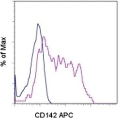
- Experimental details
- Normal human peripheral blood cells were either left unstimulated (blue histogram) or stimulated for 4 hours with LPS (purple histogram) and then stained with Anti-Human CD142 APC. Total viable cells were used for analysis.
