Explore
 Validate
 Learn
Learn Western blot
 Immunoprecipitation
Antibody data
- Antibody Data
- Antigen structure
- References [4]
- Comments [0]
- Validations
- Western blot [4]
- Immunocytochemistry [2]
- Other assay [6]
Submit
Validation data
Reference
Comment
Report error
- Product number
- MA1-21516 - Provider product page

- Provider
- Invitrogen Antibodies
- Product name
- TBP Monoclonal Antibody (1TBP18)
- Antibody type
- Monoclonal
- Antigen
- Recombinant full-length protein
- Description
- This antibody does not cross-react with Drosophila melanogaster, S. cerevisiae, silk worm, or Xenopus laevis. Store product as a concentrated solution. Centrifuge briefly prior to opening the vial.
- Reactivity
- Human, Mouse, Rat
- Host
- Mouse
- Isotype
- IgG
- Antibody clone number
- 1TBP18
- Vial size
- 100 μg
- Concentration
- 0.70 mg/mL
- Storage
- Store at 4°C short term. For long term storage, store at -20°C, avoiding freeze/thaw cycles.
Submitted references Chromatin accessibility underlies synthetic lethality of SWI/SNF subunits in ARID1A-mutant cancers.
Loss of Runx2 sensitises osteosarcoma to chemotherapy-induced apoptosis.
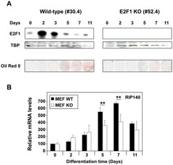
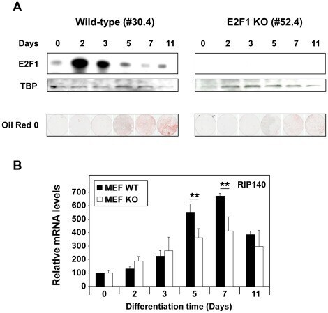
The RIP140 gene is a transcriptional target of E2F1.
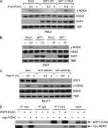
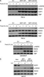
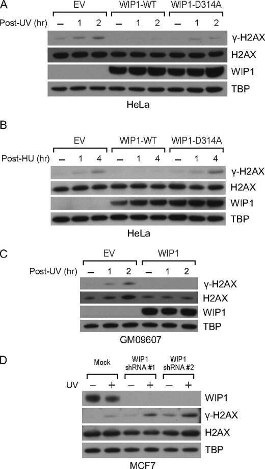
Wild-type p53-induced phosphatase 1 dephosphorylates histone variant gamma-H2AX and suppresses DNA double strand break repair.
Kelso TWR, Porter DK, Amaral ML, Shokhirev MN, Benner C, Hargreaves DC
eLife 2017 Oct 2;6
eLife 2017 Oct 2;6
Loss of Runx2 sensitises osteosarcoma to chemotherapy-induced apoptosis.
Roos A, Satterfield L, Zhao S, Fuja D, Shuck R, Hicks MJ, Donehower LA, Yustein JT
British journal of cancer 2015 Nov 3;113(9):1289-97
British journal of cancer 2015 Nov 3;113(9):1289-97
The RIP140 gene is a transcriptional target of E2F1.
Docquier A, Augereau P, Lapierre M, Harmand PO, Badia E, Annicotte JS, Fajas L, Cavaillès V
PloS one 2012;7(5):e35839
PloS one 2012;7(5):e35839
Wild-type p53-induced phosphatase 1 dephosphorylates histone variant gamma-H2AX and suppresses DNA double strand break repair.
Moon SH, Lin L, Zhang X, Nguyen TA, Darlington Y, Waldman AS, Lu X, Donehower LA
The Journal of biological chemistry 2010 Apr 23;285(17):12935-47
The Journal of biological chemistry 2010 Apr 23;285(17):12935-47
No comments: Submit comment
Supportive validation
- Submitted by
- Invitrogen Antibodies (provider)
- Main image
- Experimental details
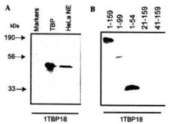
- Western Blot analysis of ecombinant human TATA Binding Protein, HeLa cell nuclear extract (A), and TBP protein fragment lysate (B) using TBP Monoclonal Antibody (1TBP18) (Product # MA1-21516). Dilution: 2 µg/mL.

- Submitted by
- Invitrogen Antibodies (provider)
- Main image

- Experimental details
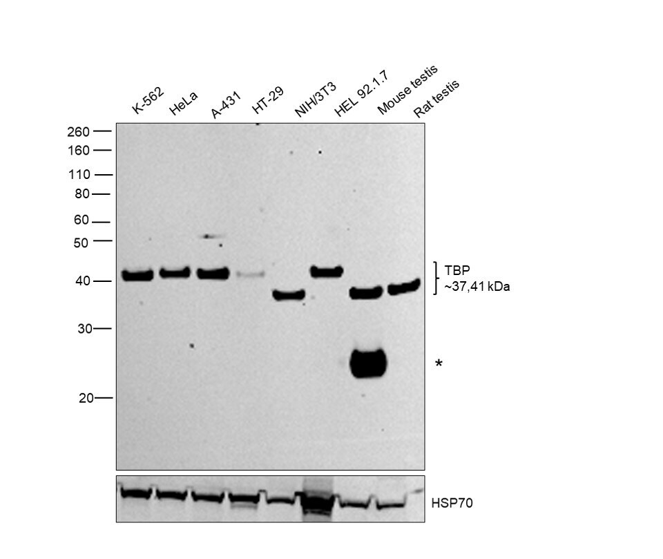
- Western blot was performed using Anti-TBP Monoclonal Antibody (1TBP18) (Product # MA1-21516) and a 41kDa band corresponding to TBP was observed across all cell lines tested, except NIH/3T3 and mouse and rat testes tissues where a 37kDa band was observed. A ~25kDa band (*) corresponding to IgG was observed in mouse testis sample. Nuclear enriched extracts (30 µg lysate) of K-562 (Lane 1), HeLa (Lane 2), A-431 (Lane 3), HT-29 (Lane 4), NIH/3T3 (Lane 5), HEL 92.1.7 (Lane 6), Mouse Testis (Lane 7), Rat Testis (Lane 8 were electrophoresed using NuPAGE™ 10% Bis-Tris Protein Gel (Product # NP0302BOX). Resolved proteins were then transferred onto a Nitrocellulose membrane (Product # IB23001) by iBlot® 2 Dry Blotting System (Product # IB21001). The blot was probed with the primary antibody (2 µg/mL concentration) and detected by chemiluminescence with Goat anti-Mouse IgG (H+L) Superclonal™ Recombinant Secondary Antibody, HRP (Product # A28177, 1:4000 dilution) using the iBright FL 1000 (Product # A32752). Chemiluminescent detection was performed using SuperSignal™ West Dura Extended Duration Substrate (Product # 34076).
- Submitted by
- Invitrogen Antibodies (provider)
- Main image
- Experimental details
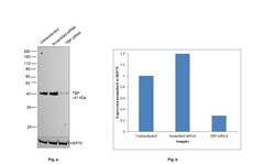
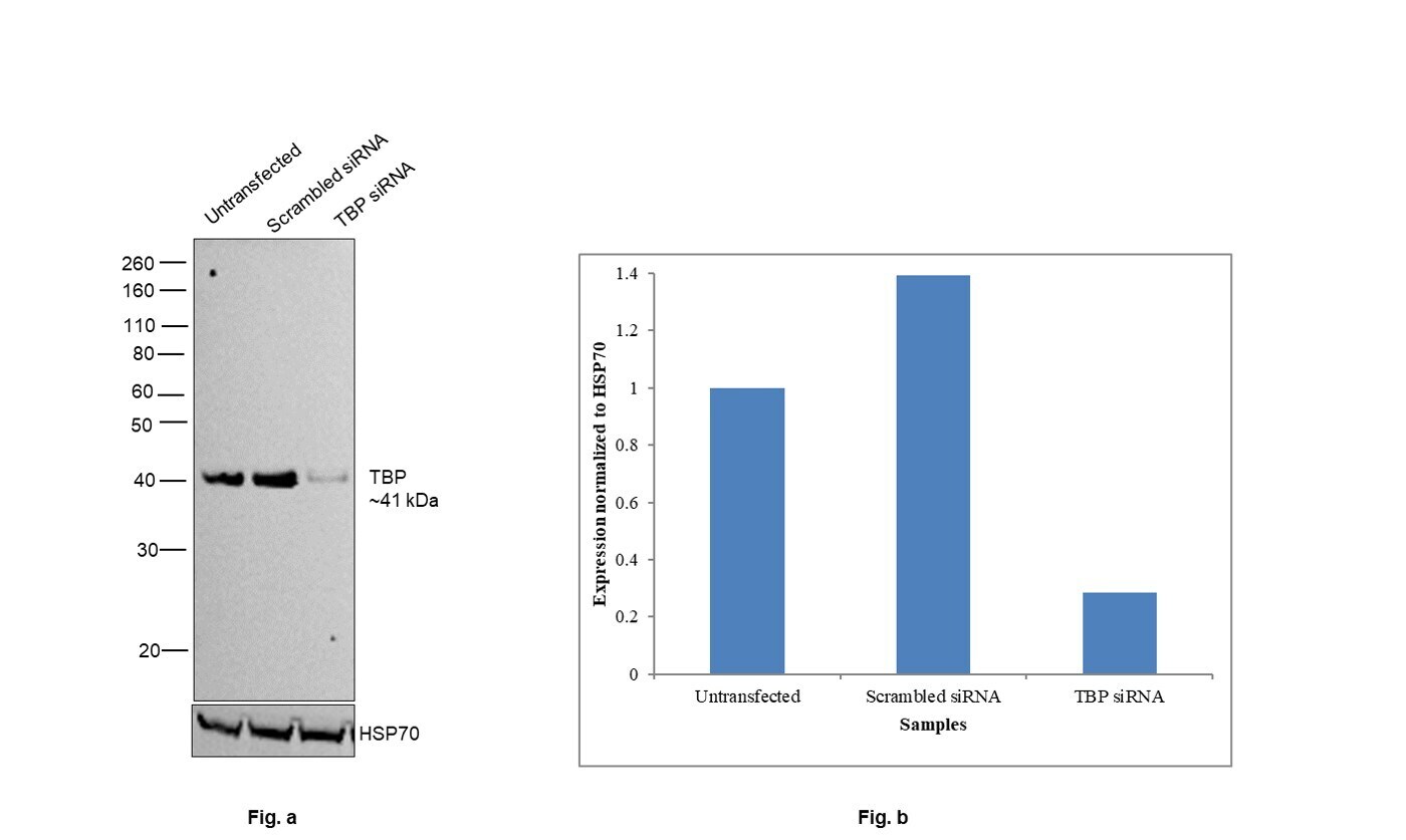
- Knockdown of TBP was achieved by transfecting HeLa with TBP specific siRNAs (Silencer® select Product # s13826, s13827). Western blot analysis (Fig. a) was performed using nuclear enriched extracts from the TBP knockdown cells (lane 3), non-targeting scrambled siRNA transfected cells (lane 2) and untransfected cells (lane 1). The blot was probed with TBP Monoclonal Antibody (1TBP18) (Product # MA1-21516, 2 µg/mL concentration) and Goat anti-Mouse IgG (H+L) Superclonal™ Recombinant Secondary Antibody, HRP (Product # A28177, 1:4000 dilution). Densitometric analysis of this western blot is shown in the histogram (Fig. b). Decrease in signal upon siRNA mediated knockdown confirms that antibody is specific to TBP.
- Submitted by
- Invitrogen Antibodies (provider)
- Main image
- Experimental details
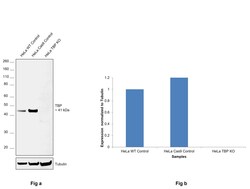
- Knockout of TBP was achieved by CRISPR-Cas9 genome editing using LentiArray™ Lentiviral sgRNA (Product # A32042, Assay ID CRISPR639612_LV) and LentiArray Cas9 Lentivirus (Product # A32064). Western blot analysis of TBP was performed by loading 30 µg of HeLa wild type (Lane 1), HeLa Cas9 (Lane 2) andHeLa TBP KO (Lane 3) modified whole cell extracts. The samples were electrophoresed using NuPAGE™ Novex™ 4-12% Bis-Tris Protein Gel (Product # NP0321BOX). Resolved proteins were then transferred onto a nitrocellulose membrane (Product # IB23001) by iBlot® 2 Dry Blotting System (Product # IB21001). The blot was probed with TBP Monoclonal Antibody (1TBP18) (Product # MA1-21516, 1:500 dilution) and Goat anti-Mouse IgG (H+L) Superclonal™ Recombinant Secondary Antibody, HRP (Product # A28177, 1:5000 dilution) using the iBright™ FL 1500 (Product # A44115). Chemiluminescent detection was performed using Novex® ECL Chemiluminescent Substrate Reagent Kit (Product # WP20005). Loss of signal upon CRISPR mediated knockout (KO) using the LentiArray™ CRISPR product line confirms that antibody is specific to TBP.

Supportive validation
- Submitted by
- Invitrogen Antibodies (provider)
- Main image

- Experimental details
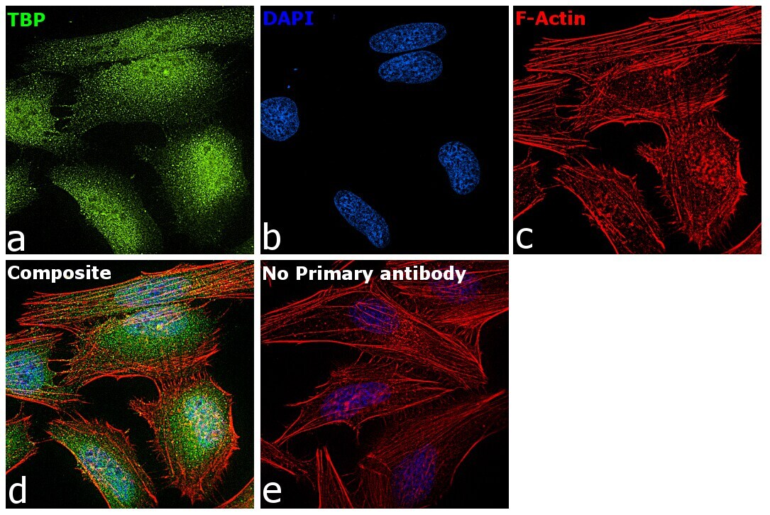
- Immunofluorescence analysis of TBP was performed using 70% confluent log phase HeLa cells. The cells were fixed with 4% paraformaldehyde for 10 minutes, permeabilized with 0.1% Triton™ X-100 for 15 minutes, and blocked with 2% BSA for 45 minutes at room temperature. The cells were labeled with TBP Monoclonal Antibody (1TBP18) (Product # MA1-21516) at 1:100 dilution in 0.1% BSA, incubated at 4 degree celsius overnight and then labeled with Donkey anti-Mouse IgG (H+L) Highly Cross-Adsorbed Secondary Antibody, Alexa Fluor Plus 488 (Product # A32766), (1:2000 dilution), for 45 minutes at room temperature (Panel a: Green). Nuclei (Panel b:Blue) were stained with ProLong™ Diamond Antifade Mountant with DAPI (Product # P36962). F-actin (Panel c: Red) was stained with Rhodamine Phalloidin (Product # R415, 1:300 dilution). Panel d represents the merged image showing predominant nuclear localization. Panel e represents control cells with no primary antibody to assess background. The images were captured at 60X magnification.

- Submitted by
- Invitrogen Antibodies (provider)
- Main image

- Experimental details
- Immunofluorescence analysis of TBP was performed using 70% confluent log phase HeLa cells. The cells were fixed with 4% paraformaldehyde for 10 minutes, permeabilized with 0.1% Triton™ X-100 for 15 minutes, and blocked with 2% BSA for 45 minutes at room temperature. The cells were labeled with TBP Monoclonal Antibody (1TBP18) (Product # MA1-21516) at 1:100 dilution in 0.1% BSA, incubated at 4 degree celsius overnight and then labeled with Donkey anti-Mouse IgG (H+L) Highly Cross-Adsorbed Secondary Antibody, Alexa Fluor Plus 488 (Product # A32766), (1:2000 dilution), for 45 minutes at room temperature (Panel a: Green). Nuclei (Panel b:Blue) were stained with ProLong™ Diamond Antifade Mountant with DAPI (Product # P36962). F-actin (Panel c: Red) was stained with Rhodamine Phalloidin (Product # R415, 1:300 dilution). Panel d represents the merged image showing predominant nuclear localization. Panel e represents control cells with no primary antibody to assess background. The images were captured at 60X magnification.
Supportive validation
- Submitted by
- Invitrogen Antibodies (provider)
- Main image
- Experimental details
- NULL

- Submitted by
- Invitrogen Antibodies (provider)
- Main image
- Experimental details
- NULL
- Submitted by
- Invitrogen Antibodies (provider)
- Main image
- Experimental details
- Figure 7 Regulation of RIP140 mRNA levels in E2F1 knock-out mice. ( A ) Adipocyte differentiation of mouse embryo fibroblasts. At different times of adipocyte differentiation, lipid accumulation was measured by Oil Red O staining in wild-type (WT) and E2F1 -/- MEFs. The levels of E2F1 protein were detected by western-blot analysis as described in Materials and Methods and loading control was performed using an anti-TBP antibody. Time 0 corresponds to the addition of the differentiation medium. ( B ) Analysis of RIP140 mRNA expression by RT-qPCR in wild-type (WT) and E2F1 -/- MEFs. Quantification were performed at the same times of adipocyte differentiation as in panel A. Data are expressed as percent of the values obtained at day 0. Statistical analysis was performed using the Student t test (**p
- Submitted by
- Invitrogen Antibodies (provider)
- Main image
- Experimental details
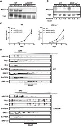
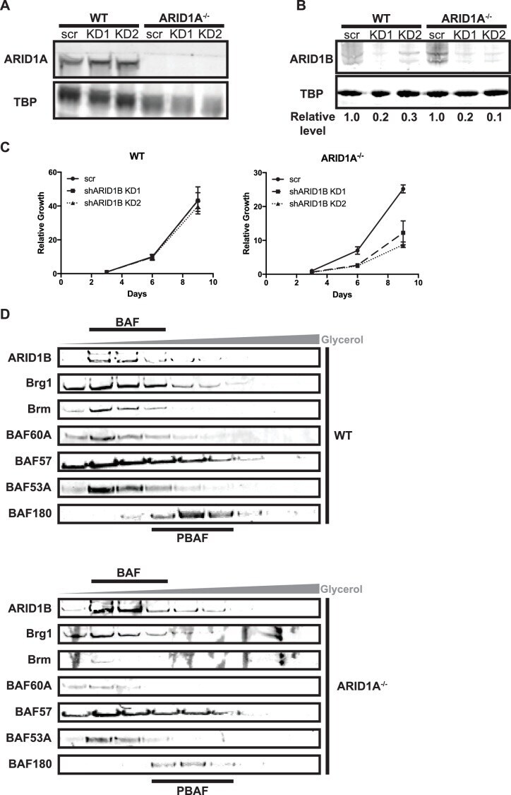
- Figure 1--figure supplement 1. Knockdown of ARID1B is synthetically lethal with ARID1A deletion in HCT116 cells. ( A ) Representative Western blot of ARID1A expression in WT and ARID1A -/- HCT116 cells with shRNAs to scrambled control (scr) or ARID1B (KD1, KD2). TBP used as loading control. Three independent experiments were performed. ( B ) Representative western blot of ARID1B expression in WT and ARID1A -/- HCT116 cells with shRNAs to scrambled control (scr) or ARID1B (KD1, KD2). Relative level indicates ARID1B expression normalized to TBP loading control. Average relative level from three independent experiments is shown in Figure 1C . ( C ) CellTiterGlo measurements after 3, 6, or 9 days of growth of WT or ARID1A -/- HCT116 cells expressing shRNAs to scrambled control (scr) or ARID1B (shARID1B KD1, shARID1B KD2) normalized to WT scr control at day 3. Averages shown from two independent biological replicates with standard deviation. ( D ) Western blot of WT and ARID1A -/- HCT116 nuclear extracts separated by glycerol gradient centrifugation. Antibodies used for blotting are indicated. Representative of three biological replicates.
- Submitted by
- Invitrogen Antibodies (provider)
- Main image
- Experimental details
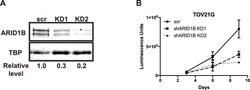
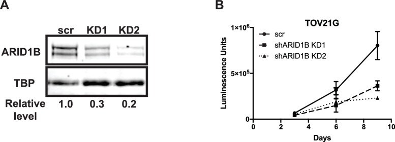
- Figure 7--figure supplement 1. Knockdown of ARID1B is synthetically lethal with ARID1A mutation in ovarian clear cell carcinoma. ( A ) Western blot of ARID1B expression in TOV21G cells with shRNAs to scrambled control (scr) or ARID1B (KD1, KD2). TBP used as loading control. Representative of two independent biological replicates. ( B ) CellTiterGlo measurements at 3, 6, or 9 days of growth of TOV21G cells expressing shRNAs to scrambled control (scr) or ARID1B (shARID1B KD1, shARID1B KD2). Averages shown from two independent biological replicates with standard deviation.
- Submitted by
- Invitrogen Antibodies (provider)
- Main image
- Experimental details
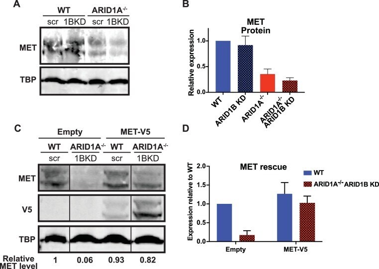
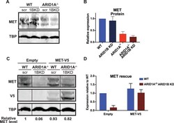
- Figure 5--figure supplement 4. MET protein expression is ARID1A/1B-dependent in HCT116 cells, but can be reconstituted by forced expression of MET. ( A ) Western blot of MET expression in WT and ARID1A-/- cells expressing scrambled or ARID1B shRNA. ( B ) MET protein expression determined by western blotting in WT and ARID1A-/- cells expressing scrambled or ARID1B shRNA. Average relative expression from three independent experiments. ( C ) Western blot of MET and V5 expression in WT and ARID1A-/- cells expressing scrambled or ARID1B shRNA and pLX_Empty or pLX_MET-V5. Representative of three independent experiments from three separate infections. ( D ) MET protein expression determined by western blotting in WT and ARID1A-/- cells expressing scrambled or ARID1B shRNA and pLX_Empty or pLX_MET-V5. Average relative expression from three independent experiments from three separate infections.