 Explore
 Validate
 Learn
Learn Western blot
Antibody data
- Antibody Data
- Antigen structure
- References [0]
- Comments [0]
- Validations
- Western blot [2]
- Immunocytochemistry [2]
- Immunohistochemistry [2]
Submit
Validation data
Reference
Comment
Report error
- Product number
- PA5-78108 - Provider product page

- Provider
- Invitrogen Antibodies
- Product name
- PLCB2 Polyclonal Antibody
- Antibody type
- Polyclonal
- Antigen
- Recombinant full-length protein
- Description
- Positive Control: THP-1, HL-60 Predicted Reactivity: Rhesus Monkey (92%) Store product as a concentrated solution. Centrifuge briefly prior to opening the vial.
- Reactivity
- Human
- Host
- Rabbit
- Isotype
- IgG
- Vial size
- 100 µL
- Concentration
- 0.71 mg/mL
- Storage
- Store at 4°C short term. For long term storage, store at -20°C, avoiding freeze/thaw cycles.
No comments: Submit comment
Supportive validation
- Submitted by
- Invitrogen Antibodies (provider)
- Main image

- Experimental details
- Western blot was performed using Anti- PLCB2 Rabbit Polyclonal Antibody (Product # PA5-78108) and a 134 kDa band corresponding to PLCB2 was observed across the cell lines tested. Whole cell extracts (30ug) of THP-1 (Lane 1), K-562 (Lane 2) and MCF7 (Lane 3) were electrophoresed using Novex® NuPAGE® 4-12 % Bis-Tris gel (Product # NP0322BOX). Resolved proteins were then transferred onto a nitrocellulose membrane (Product # IB23001) by iBlot® 2 Dry Blotting System (Product # IB21001). The blot was probed with the primary antibody (1:1000 dilution) and detected by chemiluminescence with Goat anti-Rabbit IgG (H+L), Superclonal™ Recombinant Secondary Antibody, HRP (Product # A27036, 1:4000 dilution) using the iBright FL 1000 (Product # A32752). Chemiluminescent detection was performed using Novex® ECL Chemiluminescent Substrate Reagent Kit (Product # WP20005).

- Submitted by
- Invitrogen Antibodies (provider)
- Main image

- Experimental details
- Western blot was performed using Anti-PLCB2 Polyclonal Antibody (Product # PA5-78108) and a 134 kDa band corresponding to PLCB2 was observed across cell lines. Whole cell extracts (60 µg lysate) of THP-1 (Lane 1), HEL 92.1.7 (Lane 2), NK-92 (Lane 3), Jurkat (Lane 4), HL-60 (Lane 5), U-937 (Lane 6), HeLa (Lane 7), SK-O-V3 (Lane 8) and A549 (Lane 9) were electrophoresed using NuPAGE™ 3-8% Tris-Acetate Protein Gel (Product # EA0378BOX). Resolved proteins were then transferred onto a nitrocellulose membrane (Product # IB23002) by iBlot® 2 Dry Blotting System (Product # IB21001). The blot was probed with the primary antibody (1:2000 dilution) and detected by chemiluminescence with Goat anti-Rabbit IgG (H+L) Superclonal™ Recombinant Secondary Antibody, HRP (Product # A27036,1:20000 dilution) using the iBright™ FL1500 Imaging System (Product # A44115). Chemiluminescent detection was performed using SuperSignal™ West Pico PLUS Chemiluminescent Substrate (Product # 34580). THP-1, HEL 92.17, NK-92 are high expressing than Jurkat, HL-60, U-937, HeLa, SK-O-V3 and A549 cell lines for PLCB2.

Supportive validation
- Submitted by
- Invitrogen Antibodies (provider)
- Main image

- Experimental details
- Immunocytochemistry-Immunofluorescence analysis of PLCB2 was performed in THP-1 cells fixed in 4% paraformaldehyde at RT for 15 min. Green: PLCB2 Polyclonal Antibody (Product # PA5-78108) diluted at 1:500. Blue: Fluoroshield with DAPI.

- Submitted by
- Invitrogen Antibodies (provider)
- Main image
- Experimental details
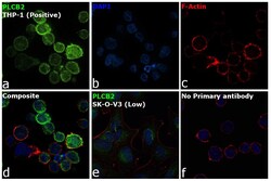
- Immunofluorescence analysis of PLCB2 was performed using 70% confluent log phase THP-1 cells. The cells were fixed with 4% paraformaldehyde for 10 minutes, permeabilized with 0.1% Triton™ X-100 for 15 minutes, and blocked with 2% BSA for 1 hour at room temperature. The cells were labeled with PLCB2 Polyclonal Antibody (Product # PA5-78108) at 1:100 dilution in 0.1% BSA, incubated at 4 degree celsius overnight and then labeled with Donkey anti-Rabbit IgG (H+L) Highly Cross-Adsorbed Secondary Antibody, Alexa Fluor Plus 488 (Product # A32790), (1:2000 dilution), for 45 minutes at room temperature (Panel a: Green). Nuclei (Panel b:Blue) were stained with ProLong™ Diamond Antifade Mountant with DAPI (Product # P36962). F-actin (Panel c: Red) was stained with Rhodamine Phalloidin (Product # R415, 1:300 dilution). Panel d represents the merged image showing cell membrane and cytosolic localization. Panel e represents SK-O-V3 cells with low expression. Panel f represents control cells with no primary antibody to assess background. The images were captured at 60X with oil immersion magnification.

Supportive validation
- Submitted by
- Invitrogen Antibodies (provider)
- Main image
- Experimental details
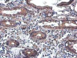
- Immunohistochemistry (Paraffin) analysis of PLCB2 was performed in paraffin-embedded human colon cancer tissue using PLCB2 Polyclonal Antibody (Product # PA5-78108) at a dilution of 1:500.

- Submitted by
- Invitrogen Antibodies (provider)
- Main image
- Experimental details
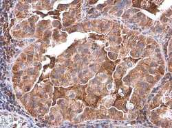
- Immunohistochemistry (Paraffin) analysis of PLCB2 was performed in paraffin-embedded human lung cancer tissue using PLCB2 Polyclonal Antibody (Product # PA5-78108) at a dilution of 1:500.
