 Explore
 Validate
 Learn
Learn Western blot
Antibody data
- Antibody Data
- Antigen structure
- References [0]
- Comments [0]
- Validations
- Western blot [3]
- Immunocytochemistry [4]
- Immunohistochemistry [2]
Submit
Validation data
Reference
Comment
Report error
- Product number
- PA5-102006 - Provider product page

- Provider
- Invitrogen Antibodies
- Product name
- PLCB2 Polyclonal Antibody
- Antibody type
- Polyclonal
- Antigen
- Synthetic peptide
- Description
- Antibody detects endogenous levels of total PLCB2.
- Reactivity
- Human, Mouse, Rat
- Host
- Rabbit
- Isotype
- IgG
- Vial size
- 100 μL
- Concentration
- 1 mg/mL
- Storage
- -20°C
No comments: Submit comment
Supportive validation
- Submitted by
- Invitrogen Antibodies (provider)
- Main image
- Experimental details
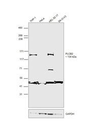
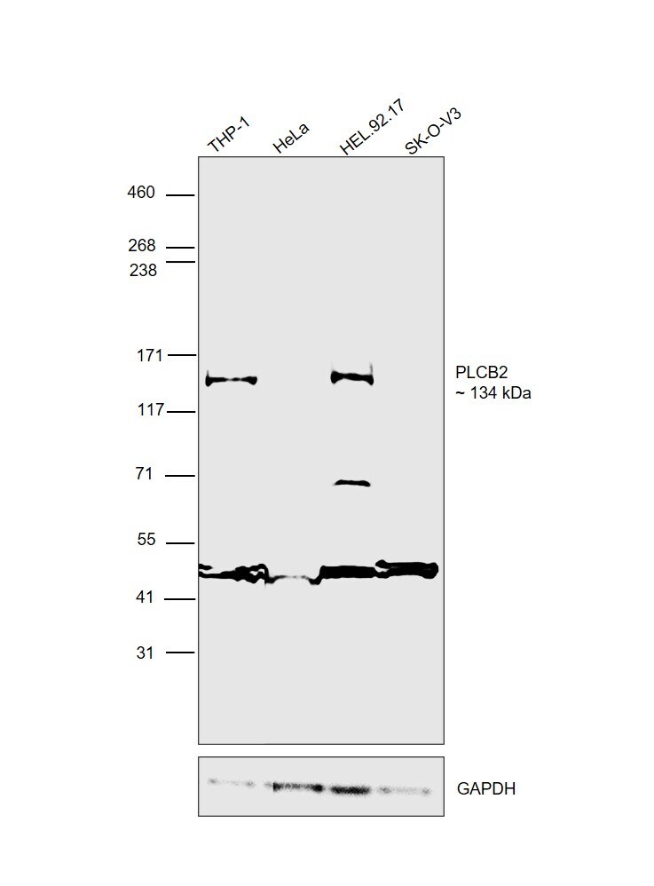
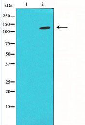
- Western blot was performed using Anti-PLCB2 Polyclonal Antibody (Product # PA5-102006) and a 134 kDa band corresponding to PLCB2 was observed across cell lines. Whole cell extracts (60 µg lysate) of THP-1 (Lane 1), HeLa (Lane 2), HEL 92.1.7 (Lane 3), SK-O-V3 (Lane 4) were electrophoresed using NuPAGE™ 3-8% Tris-Acetate Protein Gel (Product # EA0378BOX). Resolved proteins were then transferred onto a nitrocellulose membrane (Product # IB23001) by iBlot® 2 Dry Blotting System (Product # IB21001). The blot was probed with the primary antibody (1:2000 dilution) and detected by chemiluminescence with Goat anti-Rabbit IgG (Heavy Chain) Superclonal™ Recombinant Secondary Antibody, HRP (Product # A27036,1:20000 dilution) using the iBright™ FL1500 Imaging System (Product # A44115). Chemiluminescent detection was performed using SuperSignal™ West Atto Ultimate Sensitivity Substrate (Product # A38556). HeLa and SK-O-V3 are expected to be low expressing cell models for PLCB2 than THP-1 and HEL 92.1.7, as observed.
- Submitted by
- Invitrogen Antibodies (provider)
- Main image
- Experimental details
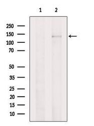
- Western blot analysis of PLCB2 in HeLa cell lysate (left lane: treated with blocking peptide). Samples were incubated with PLCB2 polyclonal antibody (Product # PA5-102006).
- Submitted by
- Invitrogen Antibodies (provider)
- Main image

- Experimental details
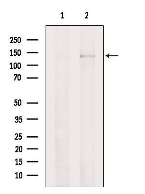
- Western blot analysis of PLCB2 in K562 cell lysate (left lane: treated with the antigen-specific peptide). Samples were incubated with PLCB2 polyclonal antibody (Product # PA5-102006).
Supportive validation
- Submitted by
- Invitrogen Antibodies (provider)
- Main image

- Experimental details
- Immunofluorescent analysis of PLCB2 in HeLa cells. Samples were fixed with paraformaldehyde, permeabilized with 0.1% Triton X-100, blocked with 10% serum (45 min at 25°C), incubated with mouse anti-beta tubulin and PLCB2 polyclonal antibody (Product # PA5-102006) using a dilution of 1:200 (1 hr, 37°C), and followed by goat anti-rabbit IgG Alexa Fluor 594 (red) and goat anti-mouse IgG Alexa Fluor 488 (green).

- Submitted by
- Invitrogen Antibodies (provider)
- Main image
- Experimental details
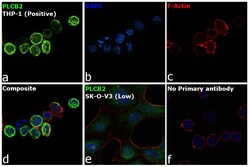
- Immunofluorescence analysis of PLCB2 was performed using 70% confluent log phase THP-1 cells. The cells were fixed with 4% paraformaldehyde for 10 minutes, permeabilized with 0.1% Triton™ X-100 for 15 minutes, and blocked with 2% BSA for 1 hour at room temperature. The cells were labeled with PLCB2 Polyclonal Antibody (Product # PA5-102006) at 1:100 dilution in 0.1% BSA, incubated at 4 degree celsius overnight and then labeled with Donkey anti-Rabbit IgG (H+L) Highly Cross-Adsorbed Secondary Antibody, Alexa Fluor Plus 488 (Product # A32790), (1:2000 dilution), for 45 minutes at room temperature (Panel a: Green). Nuclei (Panel b:Blue) were stained with ProLong™ Diamond Antifade Mountant with DAPI (Product # P36962). F-actin (Panel c: Red) was stained with Rhodamine Phalloidin (Product # R415, 1:300 dilution). Panel d represents the merged image showing cell membrane and cytosolic localization. Panel e represents control cells with no primary antibody to assess background. The images were captured at 60X with oil immersion magnification. THP-1 is high expressing than SK-O-V3 for PLCB2.
- Submitted by
- Invitrogen Antibodies (provider)
- Main image

- Experimental details
- Immunofluorescent analysis of PLCB2 in HeLa cells. Samples were fixed with paraformaldehyde, permeabilized with 0.1% Triton X-100, blocked with 10% serum (45 min at 25°C), incubated with mouse anti-beta tubulin and PLCB2 polyclonal antibody (Product # PA5-102006) using a dilution of 1:200 (1 hr, 37°C), and followed by goat anti-rabbit IgG Alexa Fluor 594 (red) and goat anti-mouse IgG Alexa Fluor 488 (green).

- Submitted by
- Invitrogen Antibodies (provider)
- Main image

- Experimental details
- Immunofluorescence analysis of PLCB2 was performed using 70% confluent log phase THP-1 cells. The cells were fixed with 4% paraformaldehyde for 10 minutes, permeabilized with 0.1% Triton™ X-100 for 15 minutes, and blocked with 2% BSA for 1 hour at room temperature. The cells were labeled with PLCB2 Polyclonal Antibody (Product # PA5-102006) at 1:100 dilution in 0.1% BSA, incubated at 4 degree celsius overnight and then labeled with Donkey anti-Rabbit IgG (H+L) Highly Cross-Adsorbed Secondary Antibody, Alexa Fluor Plus 488 (Product # A32790), (1:2000 dilution), for 45 minutes at room temperature (Panel a: Green). Nuclei (Panel b:Blue) were stained with ProLong™ Diamond Antifade Mountant with DAPI (Product # P36962). F-actin (Panel c: Red) was stained with Rhodamine Phalloidin (Product # R415, 1:300 dilution). Panel d represents the merged image showing cell membrane and cytosolic localization. Panel e represents control cells with no primary antibody to assess background. The images were captured at 60X with oil immersion magnification. THP-1 is high expressing than SK-O-V3 for PLCB2.

Supportive validation
- Submitted by
- Invitrogen Antibodies (provider)
- Main image
- Experimental details
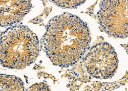
- Immunohistochemistry analysis of PLCB2 in mouse testis tissue. The sample was formaldehyde fixed and a heat mediated antigen retrieval step in citrate buffer was performed. Samples were incubated with PLCB2 polyclonal antibody (Product # PA5-102006) using a dilution of 1:100 (4°C overnight) followed by HRP conjugated anti-Rabbit secondary antibody.

- Submitted by
- Invitrogen Antibodies (provider)
- Main image

- Experimental details
- Immunohistochemistry analysis of PLCB2 in mouse testis tissue. The sample was formaldehyde fixed and a heat mediated antigen retrieval step in citrate buffer was performed. Samples were incubated with PLCB2 polyclonal antibody (Product # PA5-102006) using a dilution of 1:100 (4°C overnight) followed by HRP conjugated anti-Rabbit secondary antibody.
