Explore
 Validate
 Learn
Learn Western blot
 Immunocytochemistry
Antibody data
- Antibody Data
- Antigen structure
- References [1]
- Comments [0]
- Validations
- Immunocytochemistry [4]
- Immunohistochemistry [2]
- Other assay [1]
Submit
Validation data
Reference
Comment
Report error
- Product number
- MA5-32553 - Provider product page

- Provider
- Invitrogen Antibodies
- Product name
- GLI1 Recombinant Rabbit Monoclonal Antibody (JF09-08)
- Antibody type
- Monoclonal
- Antigen
- Synthetic peptide
- Description
- Recombinant rabbit monoclonal antibodies are produced using in vitro expression systems. The expression systems are developed by cloning in the specific antibody DNA sequences from immunoreactive rabbits. Then, individual clones are screened to select the best candidates for production. The advantages of using recombinant rabbit monoclonal antibodies include: better specificity and sensitivity, lot-to-lot consistency, animal origin-free formulations, and broader immunoreactivity to diverse targets due to larger rabbit immune repertoire.
- Reactivity
- Human, Mouse, Rat
- Host
- Rabbit
- Isotype
- IgG
- Antibody clone number
- JF09-08
- Vial size
- 100 μL
- Concentration
- 1 mg/mL
- Storage
- Store at 4°C short term. For long term storage, store at -20°C, avoiding freeze/thaw cycles.
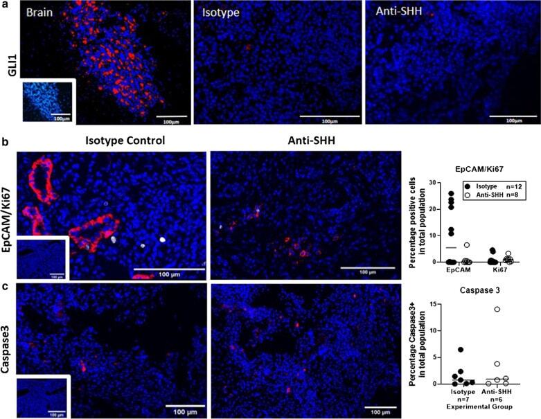
Submitted references The effects of hedgehog ligand neutralising antibody 5E1 in a mouse model of endometriosis.
Cousins FL, Farley JK, Kerrigan R, Mukherjee S, Darzi S, Gargett CE, Deane JA
BMC research notes 2020 Sep 25;13(1):454
BMC research notes 2020 Sep 25;13(1):454
No comments: Submit comment
Supportive validation
- Submitted by
- Invitrogen Antibodies (provider)
- Main image
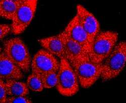
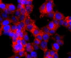
- Experimental details
- Immunocytochemical analysis of GLI1 in HepG2 cells using a GLI1 Monoclonal antibody (Product # MA5-32553) as seen in red. The nuclear counter stain is DAPI (blue). Cells were fixed in paraformaldehyde, permeabilised with 0.25% Triton X100/PBS.
- Submitted by
- Invitrogen Antibodies (provider)
- Main image

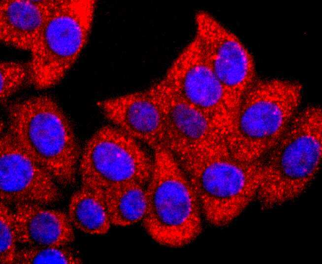
- Experimental details
- Immunocytochemical analysis of GLI1 in SH-SY-5Y cells using a GLI1 Monoclonal antibody (Product # MA5-32553) as seen in red. The nuclear counter stain is DAPI (blue). Cells were fixed in paraformaldehyde, permeabilised with 0.25% Triton X100/PBS.
- Submitted by
- Invitrogen Antibodies (provider)
- Main image
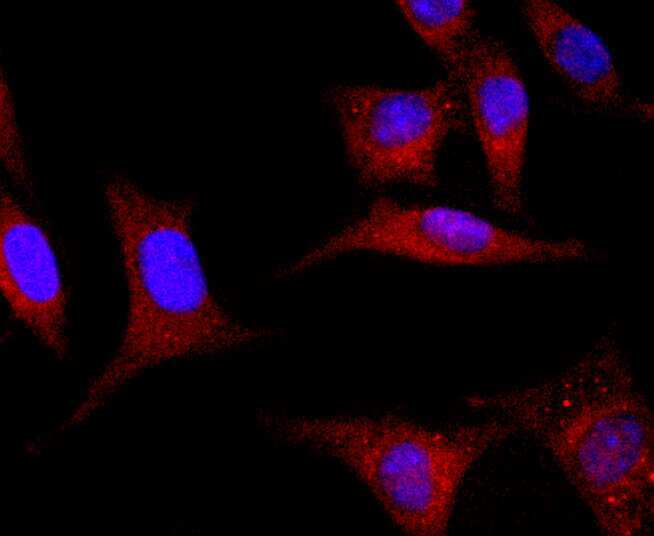
- Experimental details
- Immunocytochemical analysis of GLI1 in 293T cells using a GLI1 Monoclonal antibody (Product # MA5-32553) as seen in red. The nuclear counter stain is DAPI (blue). Cells were fixed in paraformaldehyde, permeabilised with 0.25% Triton X100/PBS.

- Submitted by
- Invitrogen Antibodies (provider)
- Main image

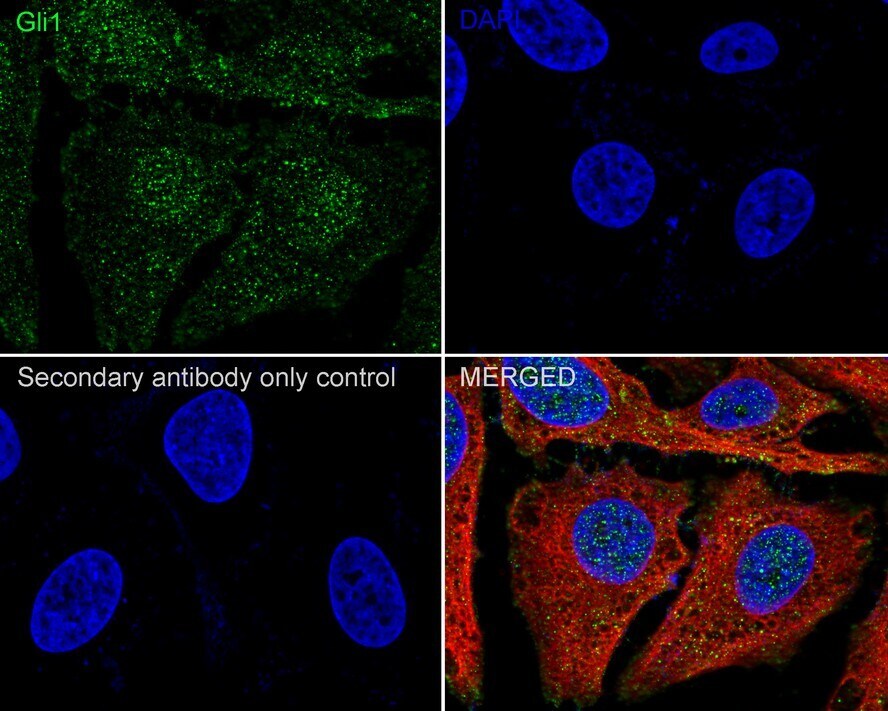
- Experimental details
- Immunocytochemistry analysis of GLI1 with HeLa cells. Cells were fixed in 4% paraformaldehyde (20 min, room temp), permeabilized with 0.1% Triton X-100 in PBS (5 min, room temp), then blocked with 1% BSA in 10% negative goat serum (1 hr, room temp), incubated with recombinant monoclonal GLI1 (Product # MA5-32553) at a dilution of 1:100 (in 1% BSA in PBST overnight at 4°C), followed by Goat Anti-Rabbit IgG H&L 488 at 1:1,000 dilution (PBS used in control). Nuclear DNA was labelled in blue with DAPI, Beta tubulin (red) at 1/100 dilution (overnight, 4°C).
Supportive validation
- Submitted by
- Invitrogen Antibodies (provider)
- Main image
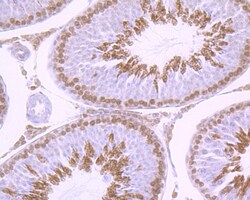
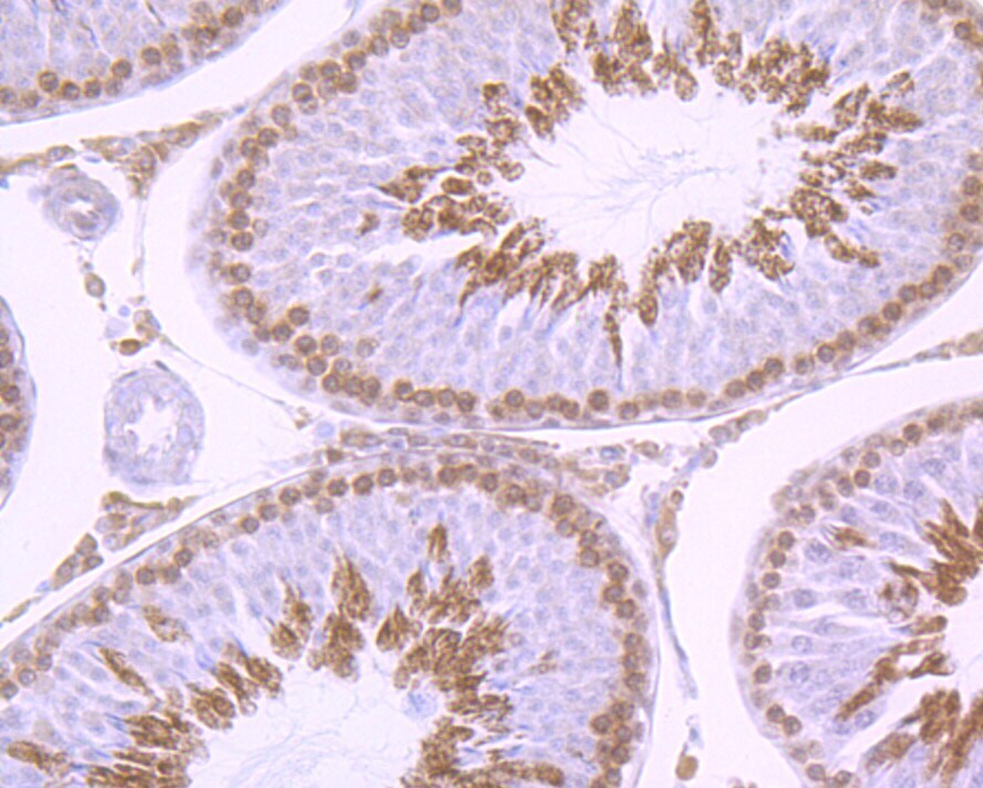
- Experimental details
- Immunohistochemical analysis of GLI1 of paraffin-embedded rat testis tissue using a GLI1 Monoclonal antibody (Product #MA5-32553). Counter stained with hematoxylin..
- Submitted by
- Invitrogen Antibodies (provider)
- Main image
- Experimental details
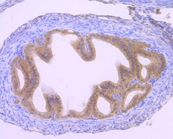
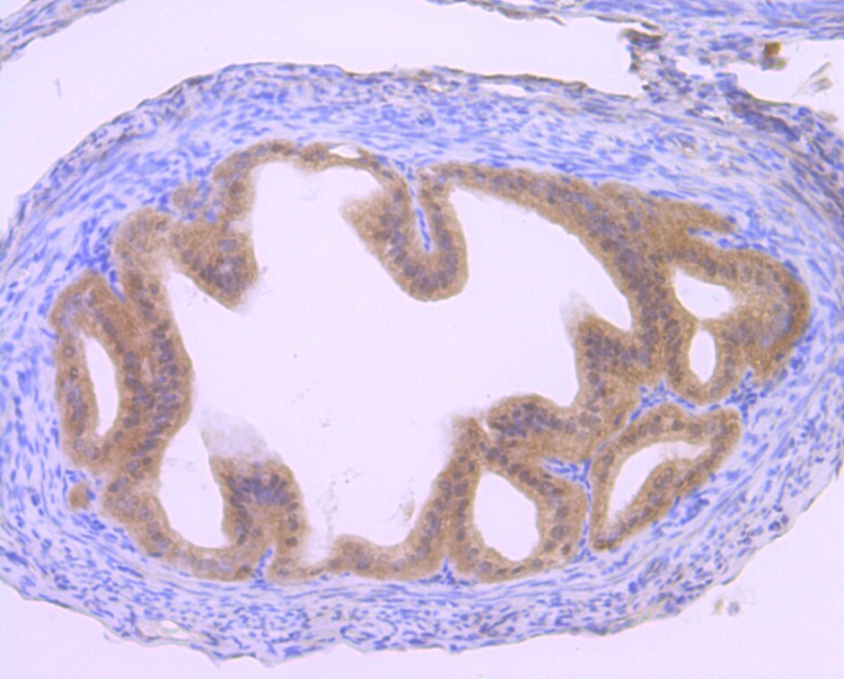
- Immunohistochemical analysis of GLI1 of paraffin-embedded Mouse fallopian tube tissue using a GLI1 Monoclonal antibody (Product #MA5-32553). Counter stained with hematoxylin..
Supportive validation
- Submitted by
- Invitrogen Antibodies (provider)
- Main image
- Experimental details
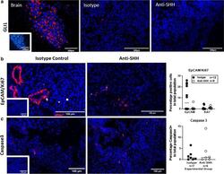
- Fig. 2 Expression of SHH pathway and cell identity markers did not change with anti-SHH treatment. a Gli1 expression (red) was detected in the mouse brain but was barely detected in lesions from either group. Inset rabbit IgG1 isotype control. b Dual immunofluorescence for EpCAM (red) and Ki67 (white), insert isotype control. c Immunofluorescence for Caspase 3 (red), insert isotype control. B + C quantification; EpCAM, Ki67 and Caspase 3 expressed as a percentage of total cell population, isotype control (black circles) and anti-Shh (white circles). Data analysed by an unpaired, two-tailed Mann-Whitney test