Explore
 Validate
 Learn
Learn Western blot
Antibody data
- Antibody Data
- Antigen structure
- References [2]
- Comments [0]
- Validations
- Western blot [1]
- Immunohistochemistry [1]
- Chromatin Immunoprecipitation [1]
Submit
Validation data
Reference
Comment
Report error
- Product number
- AF3324 - Provider product page

- Provider
- R&D Systems
- Product name
- Human GLI-1 Antibody
- Antibody type
- Polyclonal
- Description
- Antigen Affinity-purified. Detects human GLI-1 in direct ELISAs and Western blots. In Western blots, approximately 5% cross-reactivity with recombinant human GLI-3 is observed.
- Reactivity
- Human
- Host
- Goat
- Conjugate
- Unconjugated
- Antigen sequence
P08151- Isotype
- IgG
- Vial size
- 100 ug
- Concentration
- LYOPH
- Storage
- Use a manual defrost freezer and avoid repeated freeze-thaw cycles. 12 months from date of receipt, -20 to -70 °C as supplied. 1 month, 2 to 8 °C under sterile conditions after reconstitution. 6 months, -20 to -70 °C under sterile conditions after reconstitution.
Submitted references Gorlin syndrome-derived induced pluripotent stem cells are hypersensitive to hedgehog-mediated osteogenic induction.
Hedgehog and Notch signaling regulate self-renewal of undifferentiated pleomorphic sarcomas.
Hasegawa D, Ochiai-Shino H, Onodera S, Nakamura T, Saito A, Onda T, Watanabe K, Nishimura K, Ohtaka M, Nakanishi M, Kosaki K, Yamaguchi A, Shibahara T, Azuma T
PloS one 2017;12(10):e0186879
PloS one 2017;12(10):e0186879
Hedgehog and Notch signaling regulate self-renewal of undifferentiated pleomorphic sarcomas.
Wang CY, Wei Q, Han I, Sato S, Ghanbari-Azarnier R, Whetstone H, Poon R, Hu J, Zheng F, Zhang P, Wang W, Wunder JS, Alman BA
Cancer research 2012 Feb 15;72(4):1013-22
Cancer research 2012 Feb 15;72(4):1013-22
No comments: Submit comment
Supportive validation
- Submitted by
- R&D Systems (provider)
- Main image
- Experimental details
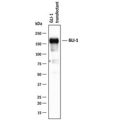
- Detection of Human GLI-1 by Western Blot. Western blot shows lysates of HEK293 human embryonic kidney cell line transfected with human GLI-1. PVDF membrane was probed with 1 µg/mL of Goat Anti-Human GLI-1 Antigen Affinity-purified Polyclonal Antibody (Catalog # AF3324) followed by HRP-conjugated Anti-Goat IgG Secondary Antibody (Catalog # HAF017). A specific band was detected for GLI-1 at approximately 165 kDa (as indicated). This experiment was conducted under reducing conditions and using Immunoblot Buffer Group 1.

Supportive validation
- Submitted by
- R&D Systems (provider)
- Main image

- Experimental details
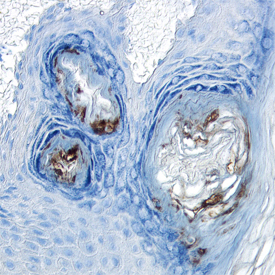
- GLI-1 in Human Skin. GLI-1 was detected in immersion fixed paraffin-embedded sections of human skin using Human GLI-1 Antigen Affinity-purified Polyclonal Antibody (Catalog # AF3324 ) at 5 µg/mL overnight at 4 °C. Tissue was stained using the Anti-Goat HRP-DAB Cell & Tissue Staining Kit (brown; Catalog # CTS008) and counterstained with hematoxylin (blue). View our protocol for Chromogenic IHC Staining of Paraffin-embedded Tissue Sections.
Supportive validation
- Submitted by
- R&D Systems (provider)
- Main image

- Experimental details
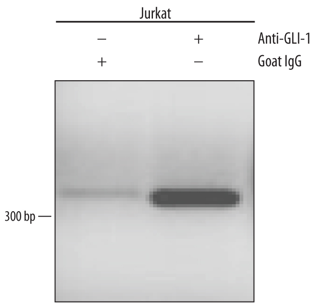
- Detection of GLI-1-regulated Genes by Chromatin Immunoprecipitation. Jurkat human acute T cell leukemia cell line treated with 50 ng/mL PMA and 200 ng/mL calcium ionomycin for 30 mintes were fixed using formaldehyde, resuspended in lysis buffer, and sonicated to shear chromatin. GLI-1/DNA complexes were immunoprecipitated using 5 μg Goat Anti-Human GLI-1 Antigen Affinity-purified Polyclonal Antibody (Catalog # AF3324) or control antibody (Catalog # AB-108-C) for 15 minutes in an ultrasonic bath, followed by Biotinylated Anti-Goat IgG Secondary Antibody (Catalog # BAF109). Immunocomplexes were captured using 50 μL of MagCellect Streptavidin Ferrofluid (Catalog # MAG999) and DNA was purified using chelating resin solution. The Bcl-2 promoter was detected by standard PCR.