Explore
 Validate
 Learn
Learn Western blot
Antibody data
- Antibody Data
- Antigen structure
- References [0]
- Comments [0]
- Validations
- Western blot [1]
- Flow cytometry [1]
Submit
Validation data
Reference
Comment
Report error
- Product number
- 10-7010-25 - Provider product page

- Provider
- ABEOMICS Inc.
- Product name
- Anti-DNMT2 Antibody
- Antibody type
- Monoclonal
- Description
- DNMT2, also known as tRNA-aspartic acid methyltransferase 1 (TRDMT1), is highly conserved cytosine-C5 methyltransferase that introduces the C38 methylation of tRNAAsp in many species, including lower eukaryotes, plants, insects, and humans. DNMT2 contains all 10 sequence motifs that are conserved among m5C MTases, including the consensus S-adenosyl-L-methionine-binding motifs and the active site ProCys dipeptide. It is a relatively small protein of 391 amino acids and lacks the large N-terminal domains present in the DNMT1 and DNMT3 families. DNMT2 is ubiquitously expressed with multiple mRNA species in most human and mouse adult tissues. It is variably expressed in human cancer cell lines.
- Reactivity
- Human, Mouse
- Host
- Mouse
- Conjugate
- Unconjugated
- Antigen sequence
A partial length recombinant protei
n from DNMT2  was used as the immun
ogen for this antibody.- Isotype
- IgG
- Antibody clone number
- ABM1H70
- Vial size
- 100 µg
- Concentration
- 0.5 mg/ml
- Storage
- Store the antibody at 4°C, stable for 6 months. For long-term storage, store at -20°C. Avoid repeat freez thawing
No comments: Submit comment
Supportive validation
- Submitted by
- ABEOMICS Inc. (provider)
- Main image

- Experimental details
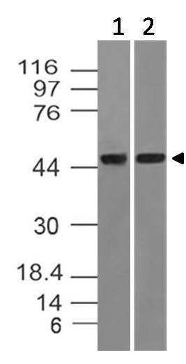
- Western blot analysis of DNMT2. Anti-DNMT2 antibody (Clone: ABM1H70) was used at 4 µg/ml on Jurkat and mouse embryonic liver lysates.
- Protocol
- Protocol
Supportive validation
- Submitted by
- ABEOMICS Inc. (provider)
- Main image
- Experimental details
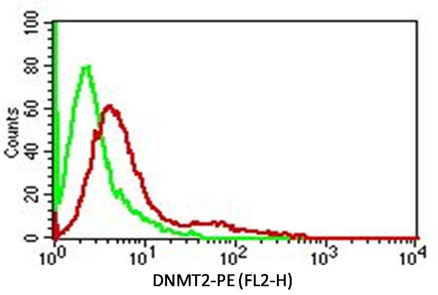
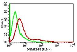
- Intracellular Flow analysis of DNMT2 antibody in Hela cells using 0.5 µg/ 10^6 cells of anti-DNMT2 antibody (ABM1H70). Green represents isotype control; red represents anti-DNMT 2 antibody. Goat anti-mouse PE conjugate was used as secondary antibody. (Cells were fixed with 4% paraformaldehyde for 10 min and washed with PBS by centrifuging at 1100 for 5 min followed by permeabilization for 20 min and washed again as mentioned above. Then cell were incubated with primary antibody for 45 min. and after washing the cells twice in PBS, incubated with conjugated secondary antibody for 30 min. Data acquisition was done after washing twice with PBS as mentioned above).
- Protocol
- Protocol