Explore
 Validate
 Learn
Learn Western blot
 ELISA
ELISAAntibody data
- Antibody Data
- Antigen structure
- References [2]
- Comments [0]
- Validations
- Western blot [1]
Submit
Validation data
Reference
Comment
Report error
- Product number
- A04950-1 - Provider product page

- Provider
- Boster Biological Technology
- Product name
- Anti-Kir6.1/KCNJ8 Antibody Picoband™
- Antibody type
- Polyclonal
- Description
- Rabbit IgG polyclonal antibody for Kir6.1/KCNJ8 detection. Tested with WB, IHC-P, FCM, Direct ELISA in Human;Mouse;Rat.
- Reactivity
- Human, Mouse, Rat
- Host
- Rabbit
- Vial size
- 100μg/vial
- Concentration
- Add 0.2ml of distilled water will yield a concentration of 500μg/ml.
- Storage
- At -20°C for one year. After reconstitution, at 4°C for one month. It can also be aliquoted and stored frozen at -20°C for a longer time. Avoid repeated freezing and thawing.
- Handling
- Add 0.2ml of distilled water will yield a concentration of 500μg/ml.
Submitted references Exogenous H2S promotes ion channel reconstruction to regulate colonic motility in rats with dinitrobenzene sulfonic acid-induced colitis.
Astragaloside IV regulates expression of ATP-sensitive potassium channel subunits after ischemia-reperfusion in rat ventricular cardiomyocytes.
Liu Y, Liao R, Qiang Z, Yang W, Cao J
Annals of translational medicine 2022 Jun;10(12):681
Annals of translational medicine 2022 Jun;10(12):681
Astragaloside IV regulates expression of ATP-sensitive potassium channel subunits after ischemia-reperfusion in rat ventricular cardiomyocytes.
Han XH, Liu P, Zhang YY, Zhang N, Chen FR, Cai JF
Journal of traditional Chinese medicine = Chung i tsa chih ying wen pan 2011 Dec;31(4):321-6
Journal of traditional Chinese medicine = Chung i tsa chih ying wen pan 2011 Dec;31(4):321-6
No comments: Submit comment
Supportive validation
- Submitted by
- Boster Biological Technology (provider)
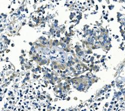
- Main image
- Experimental details
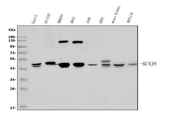
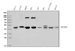
- Western blot analysis of Kir6.1/KCNJ8 using anti-Kir6.1/KCNJ8 antibody (A04950-1). Electrophoresis was performed on a 5-20% SDS-PAGE gel at 70V (Stacking gel) / 90V (Resolving gel) for 2-3 hours. The sample well of each lane was loaded with 50ug of sample under reducing conditions. Lane 1: human CACO-2 whole cell lysates, Lane 2: human SH-SY5Y whole cell lysates, Lane 3: human HEK293 whole cell lysates, Lane 4: human K562 whole cell lysates, Lane 5: human A549 whole cell lysates, Lane 6: rat RH35 whole cell lysates, Lane 7: mouse kidney tissue lysates, Lane 8: mouse HEPA1-6 whole cell lysates. After Electrophoresis, proteins were transferred to a Nitrocellulose membrane at 150mA for 50-90 minutes. Blocked the membrane with 5% Non-fat Milk/ TBS for 1.5 hour at RT. The membrane was incubated with rabbit anti-Kir6.1/KCNJ8 antigen affinity purified polyclonal antibody (Catalog # A04950-1) at 0.5 μg/mL overnight at 4°C, then washed with TBS-0.1%Tween 3 times with 5 minutes each and probed with a goat anti-rabbit IgG-HRP secondary antibody at a dilution of 1:5000 for 1.5 hour at RT. The signal is developed using an Enhanced Chemiluminescent detection (ECL) kit (Catalog # EK1002) with Tanon 5200 system. A specific band was detected for Kir6.1/KCNJ8 at approximately 48KD. The expected band size for Kir6.1/KCNJ8 is at 48KD.
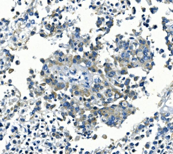
- Additional image