Explore
 Validate
 Learn
LearnA03006-1
antibody from Boster Biological Technology
Targeting: CACNA1S
CACNL1A3, Cav1.1, HOKPP, hypoPP, MHS5
 Western blot
 ELISA
ELISAAntibody data
- Antibody Data
- Antigen structure
- References [1]
- Comments [0]
- Validations
- Western blot [1]
Submit
Validation data
Reference
Comment
Report error
- Product number
- A03006-1 - Provider product page

- Provider
- Boster Biological Technology
- Product name
- Anti-CACNA1S Antibody Picoband™
- Antibody type
- Polyclonal
- Description
- Rabbit IgG polyclonal antibody for CACNA1S detection. Tested with WB, IHC-P, FCM, Direct ELISA in Human;Mouse;Rat;Monkey.
- Reactivity
- Human, Mouse, Rat, Simian
- Host
- Rabbit
- Vial size
- 100μg/vial
- Concentration
- Add 0.2ml of distilled water will yield a concentration of 500μg/ml.
- Storage
- At -20°C for one year. After reconstitution, at 4°C for one month. It can also be aliquoted and stored frozen at -20°C for a longer time. Avoid repeated freezing and thawing.
- Handling
- Add 0.2ml of distilled water will yield a concentration of 500μg/ml.
Submitted references Lactate ameliorates palmitate-induced impairment of differentiative capacity in C2C12 cells through the activation of voltage-gated calcium channels.
Wan J, Cheng C, Li X, Zhu Y, Su H, Gong Y, Ding K, Gao X, Dang C, Li G, Jiang W, Yao LH
Journal of physiology and biochemistry 2024 May;80(2):349-362
Journal of physiology and biochemistry 2024 May;80(2):349-362
No comments: Submit comment
Supportive validation
- Submitted by
- Boster Biological Technology (provider)
- Main image

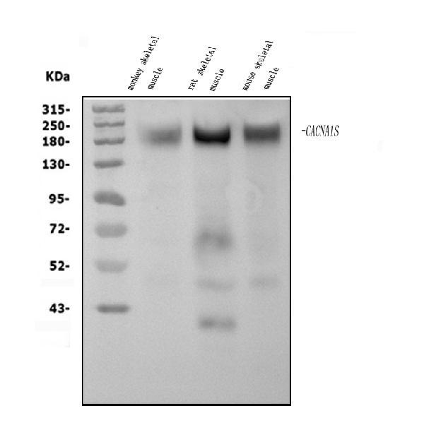
- Experimental details
- Western blot analysis of CACNA1S using anti-CACNA1S antibody (A03006-1). Electrophoresis was performed on a 5-20% SDS-PAGE gel at 70V (Stacking gel) / 90V (Resolving gel) for 2-3 hours. The sample well of each lane was loaded with 50ug of sample under reducing conditions. Lane 1: monkey skeletal muscle tissue lysates, Lane 2: rat skeletal muscle tissue lysates, Lane 3: mouse skeletal muscle tissue lysates. After Electrophoresis, proteins were transferred to a Nitrocellulose membrane at 150mA for 50-90 minutes. Blocked the membrane with 5% Non-fat Milk/ TBS for 1.5 hour at RT. The membrane was incubated with rabbit anti-CACNA1S antigen affinity purified polyclonal antibody (Catalog # A03006-1) at 0.5 μg/mL overnight at 4°C, then washed with TBS-0.1%Tween 3 times with 5 minutes each and probed with a goat anti-rabbit IgG-HRP secondary antibody at a dilution of 1:5000 for 1.5 hour at RT. The signal is developed using an Enhanced Chemiluminescent detection (ECL) kit (Catalog # EK1002) with Tanon 5200 system. A specific band was detected for CACNA1S at approximately 220KD. The expected band size for CACNA1S is at 220KD.
- Additional image