Explore
 Validate
 Learn
Learn Western blot
Antibody data
- Antibody Data
- Antigen structure
- References [0]
- Comments [0]
- Validations
- Western blot [2]
- Immunocytochemistry [1]
- Immunohistochemistry [1]
Submit
Validation data
Reference
Comment
Report error
- Product number
- PA5-102999 - Provider product page

- Provider
- Invitrogen Antibodies
- Product name
- XDH Polyclonal Antibody
- Antibody type
- Polyclonal
- Antigen
- Synthetic peptide
- Reactivity
- Human, Mouse, Rat
- Host
- Rabbit
- Isotype
- IgG
- Vial size
- 100 µL
- Concentration
- 1 mg/mL
- Storage
- -20°C
No comments: Submit comment
Supportive validation
- Submitted by
- Invitrogen Antibodies (provider)
- Main image

- Experimental details
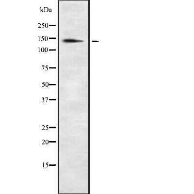
- Western blot analysis of XDH in HepG2 cell lysate. Samples were incubated with XDH polyclonal antibody (Product # PA5-102999).
- Submitted by
- Invitrogen Antibodies (provider)
- Main image
- Experimental details
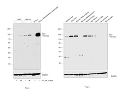
- Western blot was performed using Anti-XDH Polyclonal Antibody (Product # PA5-102999) and a 150 kDa band corresponding to XDH was upregulated in A549 and Hep G2 upon PTI treatment. The protein was upregulated in 3T3-L1 upon differentiation to adipocytes when compared to undifferentiated 3T3-L1; higher expression of protein was also observed in white adipose as compared to other tissues. Whole cell extracts (30 µg lysate) of A549 (Lane 1), A549 treated with PTI (1X, 4hrs) (Lane 2), Hep G2 (Lane 3), Hep G2 treated with PTI (1X, 4hrs) (Lane 4), 3T3-L1 (Lane 5), 3T3-L1 differentiated to adipocytes (Lane 6) in Fig. a; Mouse Liver (Lane 1), Rat Liver (Lane 2), Mouse White Adipose (Lane 3), Mouse Skeletal Muscle (Lane 4), Mouse Kidney (Lane 5), Rat Kidney (Lane 6), Mouse Testis (Lane 7), Mouse Heart (Lane 8) and Mouse Brain (Lane 9) were electrophoresed using NuPAGE™ 4-12% Bis-Tris Protein Gel (Product # NP0322BOX). Resolved proteins were then transferred onto a nitrocellulose membrane (Product # IB23001) by iBlot® 2 Dry Blotting System (Product # IB21001). The blot was probed with the primary antibody (1:1000 dilution) and detected by chemiluminescence with Goat anti-Rabbit IgG (H+L) Superclonal™ Recombinant Secondary Antibody, HRP (Product # A27036,1:20000 dilution) using the iBright™ FL1500 Imaging System (Product # A44115). Chemiluminescent detection was performed using SuperSignal™ West Pico PLUS Chemiluminescent Substrate (Product # 34580).
Supportive validation
- Submitted by
- Invitrogen Antibodies (provider)
- Main image
- Experimental details
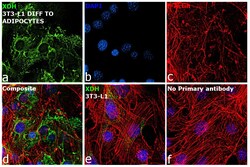
- Immunofluorescence analysis of XDH was performed using 70% confluent log phase 3T3-L1 cells and 3T3-L1 differentiated to adipocytes. The cells were fixed with 4% paraformaldehyde for 10 minutes, permeabilized with 0.1% Triton™ X-100 for 15 minutes, and blocked with 2% BSA for 45 minutes at room temperature. The cells were labeled with XDH Polyclonal Antibody (Product # PA5-102999) at 1:100 dilution in 0.1% BSA, incubated at 4 degree celsius overnight and then labeled with Donkey anti-Rabbit IgG (H+L) Highly Cross-Adsorbed Secondary Antibody, Alexa Fluor Plus 488 (Product # A32790), (1:2000 dilution), for 45 minutes at room temperature (Panel a: Green). Nuclei (Panel b:Blue) were stained with ProLong™ Diamond Antifade Mountant with DAPI (Product # P36962). F-actin (Panel c: Red) was stained with Rhodamine Phalloidin (Product # R415, 1:300). Panel d represents the merged image showing cytoplasmic localization. Panel e represents undifferentiated cells with lower expression of XDH. Panel f represents control cells with no primary antibody to assess background. The images were captured at 60X magnification.

Supportive validation
- Submitted by
- Invitrogen Antibodies (provider)
- Main image
- Experimental details
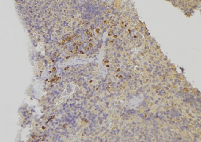
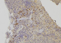
- Immunohistochemistry analysis of paraffin-embedded XDH in human lymph node tissue. Antigen retrieval was performed using citrate buffer. Samples were blocked with blocking buffer (1.5 hr, 22°C), incubated with XDH polyclonal antibody (Product # PA5-102999) using a dilution of 1:100 (1.5 hr, 22°C), followed by HRP conjugated goat anti-rabbit.