 Explore
 Validate
 Learn
Learn Western blot
 ELISA
ELISAAntibody data
- Antibody Data
- Antigen structure
- References [5]
- Comments [0]
- Validations
- Western blot [1]
- Immunocytochemistry [1]
- Immunohistochemistry [1]
Submit
Validation data
Reference
Comment
Report error
- Product number
- ABIN950569 - Provider product page

- Provider
- antibodies-online
- Product name
- anti-ATPase, H+ Transporting, Lysosomal 56/58kDa, V1 Subunit B1 (ATP6V1B1) (AA 291-318), (Middle Region) antibody
- Antibody type
- Polyclonal
- Antigen
- KLH conjugated synthetic peptide between 291-318 amino acids from the Central region of human ATP6V1B1
- Description
- Affinity chromatography on Protein A
- Reactivity
- Human
- Host
- Rabbit
- Epitope
- AA 291-318,Middle Region
- Vial size
- 0.4 mL
- Concentration
- 0.25 mg/mL
- Storage
- Store the antibody undiluted at 2-8°C for one month or (in aliquots) at -20°C for longer.
- Handling
- Avoid repeated freezing and thawing.
Submitted references Variation at the NFATC2 locus increases the risk of thiazolidinedione-induced edema in the Diabetes REduction Assessment with ramipril and rosiglitazone Medication (DREAM) study.
Distal renal tubular acidosis and its relationship with hearing loss in children: preliminary report.
Gene-centric association signals for lipids and apolipoproteins identified via the HumanCVD BeadChip.
Inner ear abnormalities in four patients with dRTA and SNHL: clinical and genetic heterogeneity.
Genetic studies in a family with distal renal tubular acidosis and sensorineural deafness.
Bailey SD, Xie C, Do R, Montpetit A, Diaz R, Mohan V, Keavney B, Yusuf S, Gerstein HC, Engert JC, Anand S, DREAM investigators
Diabetes care 2010 Oct;33(10):2250-3
Diabetes care 2010 Oct;33(10):2250-3
Distal renal tubular acidosis and its relationship with hearing loss in children: preliminary report.
Sharifian M, Esfandiar N, Mazaheri S, Kariminejad A, Mohkam M, Dalirani R, Esmaili R, Ahmadi M, Hassas-Yeganeh M
Iranian journal of kidney diseases 2010 Jul;4(3):202-6
Iranian journal of kidney diseases 2010 Jul;4(3):202-6
Gene-centric association signals for lipids and apolipoproteins identified via the HumanCVD BeadChip.
Talmud PJ, Drenos F, Shah S, Shah T, Palmen J, Verzilli C, Gaunt TR, Pallas J, Lovering R, Li K, Casas JP, Sofat R, Kumari M, Rodriguez S, Johnson T, Newhouse SJ, Dominiczak A, Samani NJ, Caulfield M, Sever P, Stanton A, Shields DC, Padmanabhan S, Melander O, Hastie C, Delles C, Ebrahim S, Marmot MG, Smith GD, Lawlor DA, Munroe PB, Day IN, Kivimaki M, Whittaker J, Humphries SE, Hingorani AD, ASCOT investigators, NORDIL investigators, BRIGHT Consortium
American journal of human genetics 2009 Nov;85(5):628-42
American journal of human genetics 2009 Nov;85(5):628-42
Inner ear abnormalities in four patients with dRTA and SNHL: clinical and genetic heterogeneity.
Andreucci E, Bianchi B, Carboni I, Lavoratti G, Mortilla M, Fonda C, Bigozzi M, Genuardi M, Giglio S, Pela I
Pediatric nephrology (Berlin, Germany) 2009 Nov;24(11):2147-53
Pediatric nephrology (Berlin, Germany) 2009 Nov;24(11):2147-53
Genetic studies in a family with distal renal tubular acidosis and sensorineural deafness.
Sethi SK, Singh N, Gil H, Bagga A
Indian pediatrics 2009 May;46(5):425-7
Indian pediatrics 2009 May;46(5):425-7
No comments: Submit comment
Supportive validation
- Submitted by
- antibodies-online (provider)
- Main image
- Experimental details
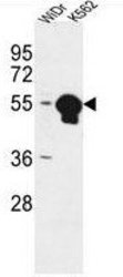
- ATP6V1B1 Antibody (Center) (Cat. #AP50306PU-N) western blot analysis in WiDr,K562 cell line lysates (35μg/lane).This demonstrates the ATP6V1B1 antibody detected the ATP6V1B1 protein (arrow).

Supportive validation
- Submitted by
- antibodies-online (provider)
- Main image
- Experimental details
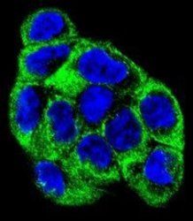
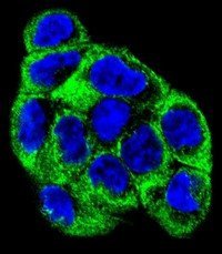
- Confocal immunofluorescent analysis of ATP6V1B1 Antibody (Center)(Cat#AP50306PU-N) with WiDr cell followed by Alexa Fluor 488-conjugated goat anti-rabbit lgG (green). DAPI was used to stain the cell nuclear (blue).
Supportive validation
- Submitted by
- antibodies-online (provider)
- Main image
- Experimental details
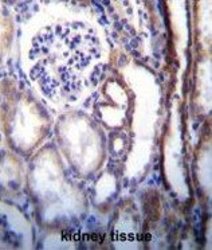
- ATP6V1B1 Antibody (Center) (Cat. #AP50306PU-N)immunohistochemistry analysis in formalin fixed and paraffin embedded human kidney tissue followed by peroxidase conjugation of the secondary antibody and DAB staining.This data demonstrates the use of ATP6V1B1 Antibody (Center) for immunohistochemistry. Clinical relevance has not been evaluated.
