Explore
 Validate
 Learn
Learn Western blot
Antibody data
- Antibody Data
- Antigen structure
- References [1]
- Comments [0]
- Validations
- Western blot [1]
- Immunohistochemistry [1]
- Other assay [1]
Submit
Validation data
Reference
Comment
Report error
- Product number
- PA5-28881 - Provider product page

- Provider
- Invitrogen Antibodies
- Product name
- T-bet Polyclonal Antibody
- Antibody type
- Polyclonal
- Antigen
- Recombinant protein fragment
- Description
- Recommended positive controls: NT2D1, IMR32, U87-MG. Predicted reactivity: Mouse (96%), Rat (97%), Pig (97%), Bovine (94%). Store product as a concentrated solution. Centrifuge briefly prior to opening the vial.
- Reactivity
- Human
- Host
- Rabbit
- Isotype
- IgG
- Vial size
- 100 µL
- Concentration
- 1 mg/mL
- Storage
- Store at 4°C short term. For long term storage, store at -20°C, avoiding freeze/thaw cycles.
Submitted references Hints on T cell responses in a fish-parasite model: Enteromyxum leei induces differential expression of T cell signature molecules depending on the organ and the infection status.
Piazzon MC, Estensoro I, Calduch-Giner JA, Del Pozo R, Picard-Sánchez A, Pérez-Sánchez J, Sitjà-Bobadilla A
Parasites & vectors 2018 Jul 31;11(1):443
Parasites & vectors 2018 Jul 31;11(1):443
No comments: Submit comment
Supportive validation
- Submitted by
- Invitrogen Antibodies (provider)
- Main image

- Experimental details
- Western Blot using T-bet Polyclonal Antibody (Product # PA5-28881). Sample (30 µg of whole cell lysate). Lane A: NT2D1. 7.5% SDS PAGE. T-bet Polyclonal Antibody (Product # PA5-28881) diluted at 1:3,000. The HRP-conjugated anti-rabbit IgG antibody was used to detect the primary antibody.

Supportive validation
- Submitted by
- Invitrogen Antibodies (provider)
- Main image
- Experimental details
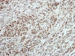
- Immunohistochemical analysis of paraffin-embedded DLD-1 xenograft, using TBX21 (Product # PA5-28881) antibody at 1:100 dilution. Antigen Retrieval: EDTA based buffer, pH 8.0, 15 min.

Supportive validation
- Submitted by
- Invitrogen Antibodies (provider)
- Main image

- Experimental details
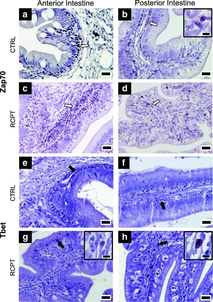
- Fig. 1 Zap70 + and Tbet + cells are more abundant in E. leei infected fish. Representative micrographs of anterior ( a , c , e , g ) and posterior ( b , d , f , h ) intestines that showed either low [control fish (CTRL): a , b , e , f ] or high [recipient fish (RCPT): c , d , g , h ] zap70 or tbet gene expression, stained with an anti-Zap70 ( a , b , c , d ) or an anti-Tbet ( e , f , g , h ) antibodies. White arrows point to some representative Zap70 + cells. Black arrows point to representative Tbet + cells. Some parasites are labeled with asterisks (*) in the posterior intestine images ( d , h ). Scale-bars : 20 mum. Insets in b , g and h show immunoreactive cells at higher magnification. Inset scale-bars : 5 mum