Explore
 Validate
 Learn
Learn Western blot
 Immunocytochemistry
 Immunohistochemistry
Antibody data
- Antibody Data
- Antigen structure
- References [1]
- Comments [0]
- Validations
- Western blot [1]
- Immunocytochemistry [1]
Submit
Validation data
Reference
Comment
Report error
- Product number
- HPA023421 - Provider product page

- Provider
- Atlas Antibodies
- Proper citation
- Atlas Antibodies Cat#HPA023421, RRID:AB_1845202
- Product name
- Anti-ATP6V1H
- Antibody type
- Polyclonal
- Description
- Polyclonal Antibody against Human ATP6V1H, Gene description: ATPase, H+ transporting, lysosomal 50/57kDa, V1 subunit H, Alternative Gene Names: CGI-11, SFD, SFDalpha, SFDbeta, VMA13, Validated applications: WB, IHC, ICC, Uniprot ID: Q9UI12, Storage: Store at +4°C for short term storage. Long time storage is recommended at -20°C.
- Reactivity
- Human
- Host
- Rabbit
- Conjugate
- Unconjugated
- Isotype
- IgG
- Vial size
- 100 µl
- Concentration
- 0.1 mg/ml
- Storage
- Store at +4°C for short term storage. Long time storage is recommended at -20°C.
- Handling
- The antibody solution should be gently mixed before use.
Submitted references Transcriptional control of autophagy–lysosome function drives pancreatic cancer metabolism
Perera R, Stoykova S, Nicolay B, Ross K, Fitamant J, Boukhali M, Lengrand J, Deshpande V, Selig M, Ferrone C, Settleman J, Stephanopoulos G, Dyson N, Zoncu R, Ramaswamy S, Haas W, Bardeesy N
Nature 2015;524(7565):361-365
Nature 2015;524(7565):361-365
No comments: Submit comment
Enhanced validation
- Submitted by
- Atlas Antibodies (provider)
- Enhanced method
- Recombinant expression validation
- Main image
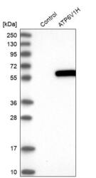
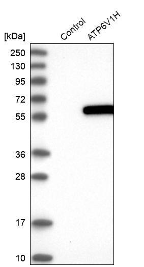
- Experimental details
- Western blot analysis in control (vector only transfected HEK293T lysate) and ATP6V1H over-expression lysate (Co-expressed with a C-terminal myc-DDK tag (~3.1 kDa) in mammalian HEK293T cells, LY414295).
- Sample type
- Human
- Protocol
- Protocol
Supportive validation
- Submitted by
- Atlas Antibodies (provider)
- Main image

- Experimental details
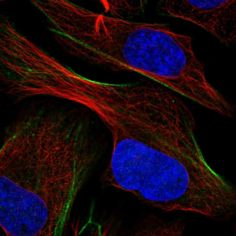
- Immunofluorescent staining of human cell line U-2 OS shows localization to actin filaments.
- Sample type
- Human