 Explore
 Validate
 Learn
Learn Western blot
Antibody data
- Antibody Data
- Antigen structure
- References [3]
- Comments [0]
- Validations
- Western blot [2]
Submit
Validation data
Reference
Comment
Report error
- Product number
- NB300-181 - Provider product page

- Provider
- Novus Biologicals
- Proper citation
- Novus Cat#NB300-181, RRID:AB_2200432
- Product name
- Rabbit Polyclonal Synapsin I Antibody
- Antibody type
- Polyclonal
- Description
- Immunogen affinity purified. Specific for ~78k synapsin I doublet protein phosphorylated at Ser603. Immunolabeling of the synapsin I band is blocked by lambda-phosphatase treatment.
- Reactivity
- Mouse, Rat, Guinea Pig
- Host
- Rabbit
- Isotype
- IgG
- Vial size
- 0.1 ml
- Storage
- Store at -20C. Avoid freeze-thaw cycles.
Submitted references Neuroprotective activities of bacopa, lycopene, astaxanthin, and vitamin B12 combination on oxidative stress-dependent neuronal death.
Long term Westernized diet leads to region-specific changes in brain signaling mechanisms.
Early Life Vitamin C Deficiency Does Not Alter Morphology of Hippocampal CA1 Pyramidal Neurons or Markers of Synaptic Plasticity in a Guinea Pig Model.
Castelli V, Melani F, Ferri C, d'Angelo M, Catanesi M, Grassi D, Benedetti E, Giordano A, Cimini A, Desideri G
Journal of cellular biochemistry 2020 May 25;
Journal of cellular biochemistry 2020 May 25;
Long term Westernized diet leads to region-specific changes in brain signaling mechanisms.
Hansen SN, Ipsen DH, Schou-Pedersen AM, Lykkesfeldt J, Tveden-Nyborg P
Neuroscience letters 2018 May 29;676:85-91
Neuroscience letters 2018 May 29;676:85-91
Early Life Vitamin C Deficiency Does Not Alter Morphology of Hippocampal CA1 Pyramidal Neurons or Markers of Synaptic Plasticity in a Guinea Pig Model.
Hansen SN, Jørgensen JMB, Nyengaard JR, Lykkesfeldt J, Tveden-Nyborg P
Nutrients 2018 Jun 8;10(6)
Nutrients 2018 Jun 8;10(6)
No comments: Submit comment
Supportive validation
- Submitted by
- Novus Biologicals (provider)
- Main image
- Experimental details
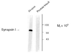
- Western Blot: Synapsin I [p Ser603] Antibody [NB300-181] - Rat cortex lysate showing specific immunolabeling of the ~78k synapsin I phosphorylated at Ser603 (Control). The phosphospecificity of this labeling is shown in the second lane (lambda-phosphatase: lambda-Ptase). The blot is identical to the control except that it was incubated in lambda-Ptase (1200 units for 30 min) before being exposed to the phospho-Ser603 synapsin I antibody. The immunolabeling is completely eliminated by treatment with lambda-Ptase.

- Submitted by
- Novus Biologicals (provider)
- Main image
- Experimental details
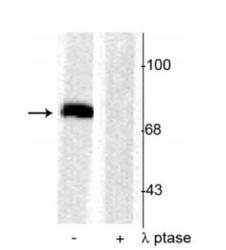
- Western Blot: Synapsin I [p Ser603] Antibody [NB300-181] - Rat cortical lysate showing specific immunolabeling of the ~78 kDa synapsin I phosphorylated at Ser603 in the first lane (-). Phosphospecificity is shown in the second lane (+) where the immunolabeling is completely eliminated by blot treatment with lambda phosphatase (l-Ptase, 1200 units for 30 minutes).
