 Explore
 Validate
 Learn
Learn Western blot
Antibody data
- Antibody Data
- Antigen structure
- References [1]
- Comments [0]
- Validations
- Western blot [2]
- Other assay [1]
Submit
Validation data
Reference
Comment
Report error
- Product number
- OPA1-04001 - Provider product page

- Provider
- Invitrogen Antibodies
- Product name
- Synapsin 1 Polyclonal Antibody
- Antibody type
- Polyclonal
- Antigen
- Purifed from natural sources
- Description
- In Western blot, this antibody detects an ~77 kDa and ~80 kDa protein representing synapsin I alpha and synapsin I beta respectively, in rat brain homogenate. Immunohistochemical staining of synapsin 1 alpha and 1 beta in human brain with OPA1-04001 yields a pattern consistent with cytoplasmic vesicle staining.
- Reactivity
- Human, Mouse, Rat, Bovine
- Host
- Rabbit
- Isotype
- IgG
- Vial size
- 50 µL
- Concentration
- Conc. Not Determined
- Storage
- -20° C, Avoid Freeze/Thaw Cycles
Submitted references Hierarchical organization and genetically separable subfamilies of PSD95 postsynaptic supercomplexes.
Frank RAW, Zhu F, Komiyama NH, Grant SGN
Journal of neurochemistry 2017 Aug;142(4):504-511
Journal of neurochemistry 2017 Aug;142(4):504-511
No comments: Submit comment
Supportive validation
- Submitted by
- Invitrogen Antibodies (provider)
- Main image

- Experimental details
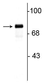
- Western blot of Synapsin 1 in rat hippocampal lysate showing specific immunolabeling of a ~78 kDa band corresponding to Synapsin 1 polyclonal antibody (Product # OPA1-04001).
- Submitted by
- Invitrogen Antibodies (provider)
- Main image
- Experimental details
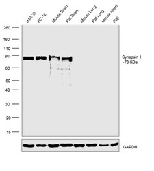
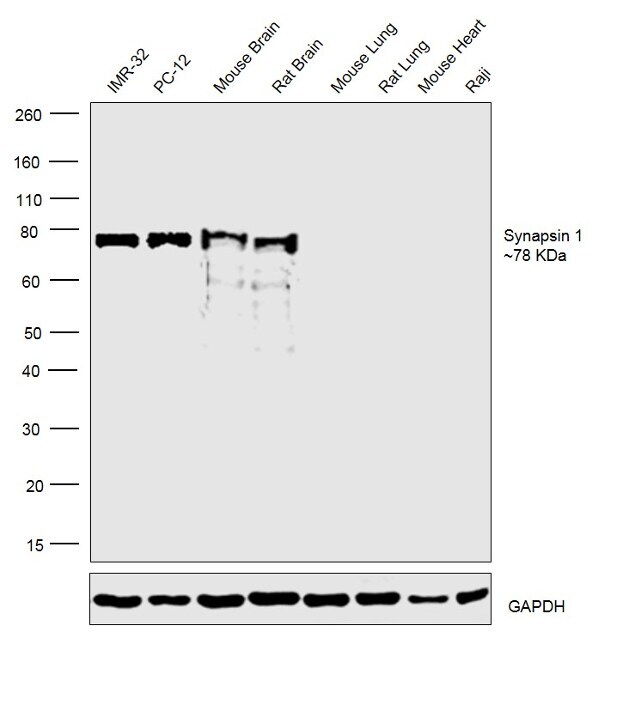
- Western blot was performed using anti-Synapsin1 Polyclonal Antibody (Product # OPA1-04001) and 78 kDa band corresponding to Synapsin1 was observed across cell lines and tissues tested except Mouse and Rat Lung, Mouse Heart and Raji which are reported to be negative. Whole cell Extracts of (30ug lysate) of IMR-32 (Lane 1), PC-12 (Lane 2), Raji (Lane 8), tissue extracts of Mouse Brain (Lane 3), Rat Brain (Lane 4), Mouse Lung (Lane 5), Rat Lung (Lane 6) and Mouse Heart (Lane 7) were electrophoresed using Novex® NuPAGE® 4-12 % Bis-Tris gel (Product # NP0322BOX). Resolved proteins were then transferred onto a nitrocellulose membrane (Product # IB23001) by iBlot® 2 Dry Blotting System (Product # IB21001).The blot was probed with the primary antibody (1:1000 dilution) and detected by chemiluminescence with Goat anti-Rabbit IgG (H+L) Superclonal™ Recombinant Secondary Antibody, HRP (Product # A27036, 1:4000 dilution). Chemiluminescent detection was performed using Novex® ECL Chemiluminescent Substrate Reagent Kit (Product # WP20005).
Supportive validation
- Submitted by
- Invitrogen Antibodies (provider)
- Main image

- Experimental details
- Figure 4 Neuroanatomically restricted assembly of Kir2.3-N-methyl D-aspartic acid receptors ( NMDAR ) ion channel-channel supercomplexes. (a) Regional enrichment of Kir2.3 expression in mouse brain. Left, mouse brain sagittal sections (lower) stained with Kir2.3 antibodies and (upper) reference Nissl stain (Allen Brain Atlas). High expression in rostroventral midbrain and caudodorsal forebrain shown and boxed regions were dissected for TAP -purification of NMDAR . White arrow indicates boxed region of piriform cortex used in high-magnification images in Fig. 4b and c. Scale bar, 1 mm. Right, BNP immunoblots of TAP -purified receptors from dissected brain regions of Glun1 TAP / TAP mice as indicated. Left immunoblot probed with Flag ( TAP -GluN1) and right immunoblot probed with Kir2.3 showing 1.5-Kir2.3- NMDAR ion channel-channel supercomplex enriched in rostroventral midbrain. Filled arrow, 1.5-Kir2.3/ NR . Molecular weight in MD a shown on right. IB , immunoblotting antibody. (b) Kir2.3 localization in layers of piriform cortex of wildtype ( WT ) and Psd95 -/- mice, including layers 1a (L1a) and 2/3 (L2/3) of piriform cortex stained with antibodies to Kir2.3 (green) and nuclear stain ( DAPI , blue). White boxes indicate regions further magnified in Fig. 4c. Scale bar, 6 mum. (c) Kir2.3 localization requires Psd95 . Higher magnification of boxed regions in Fig. 4b shows synaptic localization of Kir2.3 is disrupted in Psd95 -/- mice. Sections double-stained with Kir2.3 (gre
