 Explore
 Validate
 Learn
Learn Western blot
 Immunoprecipitation
Antibody data
- Antibody Data
- Antigen structure
- References [17]
- Comments [0]
- Validations
- Western blot [2]
- Other assay [6]
Submit
Validation data
Reference
Comment
Report error
- Product number
- 51-5200 - Provider product page

- Provider
- Invitrogen Antibodies
- Product name
- Synapsin 1 Polyclonal Antibody
- Antibody type
- Polyclonal
- Antigen
- Purifed from natural sources
- Description
- The antibody should be reconstituted in 50 uL phosphate buffered saline (PBS: 137 mM NaCl, 7.5 mM Na2HPO4, 2.7 mM KCl, 1.5 mM KH2PO4, pH 7.4) before use. After reconstitution the antibody should be aliquot and stored at -20C.
- Reactivity
- Human, Mouse, Rat, Bovine
- Host
- Rabbit
- Isotype
- IgG
- Vial size
- 10 µg
- Storage
- -20° C, Avoid Freeze/Thaw Cycles
Submitted references Intranasal Dantrolene as a Disease-Modifying Drug in Alzheimer 5XFAD Mice.
Effects of Dexamethasone on Remodeling of the Hippocampal Synaptic Filamentous Actin Cytoskeleton in a Model of Pilocarpine-induced Status Epilepticus.
Transplantation of iPS cell-derived neural progenitors overexpressing SDF-1α increases regeneration and functional recovery after ischemic stroke.
Glimepiride protects neurons against amyloid-β-induced synapse damage.
Biophysical constraints of optogenetic inhibition at presynaptic terminals.
cAMP-Inhibits Cytoplasmic Phospholipase A₂ and Protects Neurons against Amyloid-β-Induced Synapse Damage.
The progressive changes of filamentous actin cytoskeleton in the hippocampal neurons after pilocarpine-induced status epilepticus.
CCL2-ethanol interactions and hippocampal synaptic protein expression in a transgenic mouse model.
Increased astrocyte expression of IL-6 or CCL2 in transgenic mice alters levels of hippocampal and cerebellar proteins.
The rearrangement of filamentous actin in mossy fiber synapses in pentylenetetrazol-kindled C57BL/6 mice.
iPSC Transplantation increases regeneration and functional recovery after ischemic stroke in neonatal rats.
Learning-related translocation of δ-opioid receptors on ventral striatal cholinergic interneurons mediates choice between goal-directed actions.
Neurodegeneration induced by clustering of sialylated glycosylphosphatidylinositols of prion proteins.
Altered synaptic transmission in the hippocampus of transgenic mice with enhanced central nervous systems expression of interleukin-6.
Neuroadaptive changes in cerebellar neurons induced by chronic exposure to IL-6.
Spine formation and maturation in the developing rat auditory cortex.
Altered hippocampal synaptic transmission in transgenic mice with astrocyte-targeted enhanced CCL2 expression.
Shi Y, Zhang L, Gao X, Zhang J, Ben Abou M, Liang G, Meng Q, Hepner A, Eckenhoff MF, Wei H
Journal of Alzheimer's disease : JAD 2020;76(4):1375-1389
Journal of Alzheimer's disease : JAD 2020;76(4):1375-1389
Effects of Dexamethasone on Remodeling of the Hippocampal Synaptic Filamentous Actin Cytoskeleton in a Model of Pilocarpine-induced Status Epilepticus.
Yang N, Zhang Y, Wang JT, Chen C, Song Y, Liang JM, Ma DH, Zhang YF
International journal of medical sciences 2020;17(12):1683-1691
International journal of medical sciences 2020;17(12):1683-1691
Transplantation of iPS cell-derived neural progenitors overexpressing SDF-1α increases regeneration and functional recovery after ischemic stroke.
Chau M, Deveau TC, Song M, Wei ZZ, Gu X, Yu SP, Wei L
Oncotarget 2017 Nov 14;8(57):97537-97553
Oncotarget 2017 Nov 14;8(57):97537-97553
Glimepiride protects neurons against amyloid-β-induced synapse damage.
Osborne C, West E, Nolan W, McHale-Owen H, Williams A, Bate C
Neuropharmacology 2016 Feb;101:225-36
Neuropharmacology 2016 Feb;101:225-36
Biophysical constraints of optogenetic inhibition at presynaptic terminals.
Mahn M, Prigge M, Ron S, Levy R, Yizhar O
Nature neuroscience 2016 Apr;19(4):554-6
Nature neuroscience 2016 Apr;19(4):554-6
cAMP-Inhibits Cytoplasmic Phospholipase A₂ and Protects Neurons against Amyloid-β-Induced Synapse Damage.
Bate C, Williams A
Biology 2015 Sep 16;4(3):591-606
Biology 2015 Sep 16;4(3):591-606
The progressive changes of filamentous actin cytoskeleton in the hippocampal neurons after pilocarpine-induced status epilepticus.
Xiong T, Liu J, Dai G, Hou Y, Tan B, Zhang Y, Li S, Song Y, Liu H, Li Y, Li Y
Epilepsy research 2015 Dec;118:55-67
Epilepsy research 2015 Dec;118:55-67
CCL2-ethanol interactions and hippocampal synaptic protein expression in a transgenic mouse model.
Gruol DL, Vo K, Bray JG, Roberts AJ
Frontiers in integrative neuroscience 2014;8:29
Frontiers in integrative neuroscience 2014;8:29
Increased astrocyte expression of IL-6 or CCL2 in transgenic mice alters levels of hippocampal and cerebellar proteins.
Gruol DL, Vo K, Bray JG
Frontiers in cellular neuroscience 2014;8:234
Frontiers in cellular neuroscience 2014;8:234
The rearrangement of filamentous actin in mossy fiber synapses in pentylenetetrazol-kindled C57BL/6 mice.
Zhang YF, Li SL, Xiong TQ, Yang LB, Li YN, Tan BH, Liu Q, Li YC
Epilepsy research 2014 Jan;108(1):20-8
Epilepsy research 2014 Jan;108(1):20-8
iPSC Transplantation increases regeneration and functional recovery after ischemic stroke in neonatal rats.
Chau MJ, Deveau TC, Song M, Gu X, Chen D, Wei L
Stem cells (Dayton, Ohio) 2014 Dec;32(12):3075-87
Stem cells (Dayton, Ohio) 2014 Dec;32(12):3075-87
Learning-related translocation of δ-opioid receptors on ventral striatal cholinergic interneurons mediates choice between goal-directed actions.
Bertran-Gonzalez J, Laurent V, Chieng BC, Christie MJ, Balleine BW
The Journal of neuroscience : the official journal of the Society for Neuroscience 2013 Oct 9;33(41):16060-71
The Journal of neuroscience : the official journal of the Society for Neuroscience 2013 Oct 9;33(41):16060-71
Neurodegeneration induced by clustering of sialylated glycosylphosphatidylinositols of prion proteins.
Bate C, Williams A
The Journal of biological chemistry 2012 Mar 9;287(11):7935-44
The Journal of biological chemistry 2012 Mar 9;287(11):7935-44
Altered synaptic transmission in the hippocampus of transgenic mice with enhanced central nervous systems expression of interleukin-6.
Nelson TE, Olde Engberink A, Hernandez R, Puro A, Huitron-Resendiz S, Hao C, De Graan PN, Gruol DL
Brain, behavior, and immunity 2012 Aug;26(6):959-71
Brain, behavior, and immunity 2012 Aug;26(6):959-71
Neuroadaptive changes in cerebellar neurons induced by chronic exposure to IL-6.
Gruol DL, Puro A, Hao C, Blakely P, Janneke E, Vo K
Journal of neuroimmunology 2011 Oct 28;239(1-2):28-36
Journal of neuroimmunology 2011 Oct 28;239(1-2):28-36
Spine formation and maturation in the developing rat auditory cortex.
Schachtele SJ, Losh J, Dailey ME, Green SH
The Journal of comparative neurology 2011 Nov 1;519(16):3327-45
The Journal of comparative neurology 2011 Nov 1;519(16):3327-45
Altered hippocampal synaptic transmission in transgenic mice with astrocyte-targeted enhanced CCL2 expression.
Nelson TE, Hao C, Manos J, Ransohoff RM, Gruol DL
Brain, behavior, and immunity 2011 Jun;25 Suppl 1(0 1):S106-19
Brain, behavior, and immunity 2011 Jun;25 Suppl 1(0 1):S106-19
No comments: Submit comment
Supportive validation
- Submitted by
- Invitrogen Antibodies (provider)
- Main image
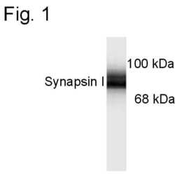
- Experimental details
- Western blot analysis of Synapsin 1 in 10 µg of rat brain lysate using Synapsin 1 polyclonal antibody (Product # 51-5200). Results show specific immunolabeling of the ~78 kDa Synapsin I doublet.

- Submitted by
- Invitrogen Antibodies (provider)
- Main image
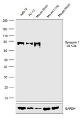
- Experimental details
- Western blot was performed using anti-Synapsin1 Polyclonal Antibody (Product # 51-5200) and 78 kDa band corresponding to Synapsin1 was observed across cell lines tested and tissues except Mouse Lung and Heart which are reported to be negative. Whole cell extracts of (30ug lysate) of IMR-32 (Lane 1), PC-12 (Lane 2), tissue extracts of Mouse Brain (Lane 3), Mouse Lung (Lane 4) and Mouse Heart (Lane 5) were electrophoresed using Novex® NuPAGE® 4-12 % Bis-Tris gel (Product # NP0322BOX). Resolved proteins were then transferred onto a nitrocellulose membrane (Product # IB23001) by iBlot® 2 Dry Blotting System (Product # IB21001).The blot was probed with the primary antibody (1:1000 dilution) and detected by chemiluminescence with Goat anti-Rabbit IgG (H+L) Superclonal™ Recombinant Secondary Antibody, HRP (Product # A27036, 1:4000 dilution).Chemiluminescent detection was performed using Novex® ECL Chemiluminescent Substrate Reagent Kit (Product # WP20005).

Supportive validation
- Submitted by
- Invitrogen Antibodies (provider)
- Main image

- Experimental details
- NULL

- Submitted by
- Invitrogen Antibodies (provider)
- Main image

- Experimental details
- NULL

- Submitted by
- Invitrogen Antibodies (provider)
- Main image

- Experimental details
- NULL

- Submitted by
- Invitrogen Antibodies (provider)
- Main image

- Experimental details
- Figure 3 Levels of synaptic proteins in CCL2-tg and non-tg hippocampus determined by Western blot. Values are mean +- SEM. Numbers in bars are the number of animals studied. Representative Western blots are shown above the respective bar graph. Top blot is the protein indicated for the graph; bottom blot (arrow) is beta actin in the same lane. Numbers in bars are the number of animals measured. n, non-tg, t, CCL2-tg. * statistically significant difference ( p < 0.05, unpaired t -test) from non-tg of the same treatment group.

- Submitted by
- Invitrogen Antibodies (provider)
- Main image

- Experimental details
- Figure 1 Abeta causes synapse damage in cultured neurons: ( A ) Immunoblots showing the amounts of synapsin-1, VAMP-1 and caveolin in neurons incubated with brain extract as shown; ( B ) The amounts of synaptophysin in neurons incubated with control medium (#), brain extract (#), Abeta-depleted brain extract (striped bar) or mock-depleted brain extract (hatched bar). Values are means +- SD from triplicate experiments performed 2 times ( n = 6). * = synaptophysin significantly less than in control neurons.

- Submitted by
- Invitrogen Antibodies (provider)
- Main image

- Experimental details
- Figure 1 Sustained activation of eArch3.0 increases presynaptic Ca 2+ and neurotransmitter release. (a) Evaluation of SyGCaMP6s targeting to the presynaptic terminal. Left: cultured neurons expressing SyGCaMP6s; center: anti-Synapsin I labeling; right: merge. (b) Representative time-averaged images acquired during the indicated time periods from a hippocampal neuron culture co-expressing SyGCaMP6s and eArch3.0. (c-d) Relative fluorecence change from baseline ( DeltaF/F ) of spontaneously active neuronal cultures expressing SyGCaMP6s. (d) Average DeltaF/F during baseline, light (590 nm, 2 mW mm -2 ) and post-light periods depicted in c. Neurons co-expressing eArch3.0 ( n = 12) showed increased SyGCaMP6s signal compared with neurons expressing eNpHR3.0 ( n = 12) and controls ( n = 8). (e) Peak SyGCaMP6s fluorescence during 590 nm illumination relative to peak SyGCaMP6s fluorescence during baseline activity of neurons expressing SyGCaMP6s (control) or co-expressing SyGCaMP6s with eArch3.0 or eNpHR3.0. Dotted line indicates zero. (f) Average EPSC rates recorded in neuronal cultures expressing the indicated constructs. Inset depicts representative voltage clamp recording traces of an eArch3.0 expressing culture before (top), during (middle) and after (bottom) 590 nm illumination (scale bar, 30 pA, 150 ms). (g) Average EPSC rates during baseline, light and post-light periods, as depicted in f (control, n = 11; eNpHR3.0, n = 12; eArch3.0, n = 12). (h) Peak EPSC rates during illumina
