Explore
 Validate
 Learn
Learn Western blot
 ELISA
ELISAAntibody data
- Antibody Data
- Antigen structure
- References [2]
- Comments [0]
- Validations
- ELISA [2]
- Immunocytochemistry [6]
- Immunohistochemistry [1]
- Flow cytometry [2]
Submit
Validation data
Reference
Comment
Report error
- Product number
- MA5-31919 - Provider product page

- Provider
- Invitrogen Antibodies
- Product name
- Synapsin 1 Monoclonal Antibody (7H10G6)
- Antibody type
- Monoclonal
- Antigen
- Purifed from natural sources
- Description
- MA5-31919 has been tested in indirect ELISA.
- Reactivity
- Human, Mouse, Rat
- Host
- Mouse
- Isotype
- IgG
- Antibody clone number
- 7H10G6
- Vial size
- 100 μL
- Concentration
- 1 mg/mL
- Storage
- Store at 4°C short term. For long term storage, store at -20°C, avoiding freeze/thaw cycles.
Submitted references SEQUIN: An imaging and analysis platform for quantification and characterization of synaptic structures in mouse.
Alzheimer's Disease-Like Neurodegeneration in Porphyromonas gingivalis Infected Neurons with Persistent Expression of Active Gingipains.
Reitz SJ, Sauerbeck AD, Kummer TT
STAR protocols 2021 Mar 19;2(1):100268
STAR protocols 2021 Mar 19;2(1):100268
Alzheimer's Disease-Like Neurodegeneration in Porphyromonas gingivalis Infected Neurons with Persistent Expression of Active Gingipains.
Haditsch U, Roth T, Rodriguez L, Hancock S, Cecere T, Nguyen M, Arastu-Kapur S, Broce S, Raha D, Lynch CC, Holsinger LJ, Dominy SS, Ermini F
Journal of Alzheimer's disease : JAD 2020;75(4):1361-1376
Journal of Alzheimer's disease : JAD 2020;75(4):1361-1376
No comments: Submit comment
Supportive validation
- Submitted by
- Invitrogen Antibodies (provider)
- Main image
- Experimental details
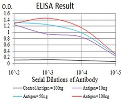
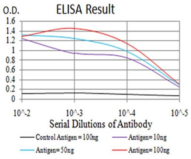
- ELISA analysis of Synapsin 1 in Control Antigen (black line, 100 ng); Antigen (purple line, 10 ng); Antigen (blue line, 50 ng); Antigen (red line, 100 ng). Samples were incubated with Synapsin 1 monoclonal antibody (Product # MA5-31919) using a dilution of 1:10,000.
- Submitted by
- Invitrogen Antibodies (provider)
- Main image

- Experimental details
- ELISA analysis of Synapsin 1 in Control Antigen (black line, 100 ng); Antigen (purple line, 10 ng); Antigen (blue line, 50 ng); Antigen (red line, 100 ng). Samples were incubated with Synapsin 1 monoclonal antibody (Product # MA5-31919) using a dilution of 1:10,000.

Supportive validation
- Submitted by
- Invitrogen Antibodies (provider)
- Main image

- Experimental details
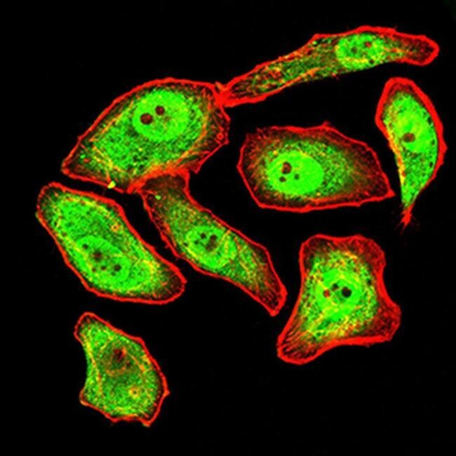
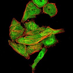
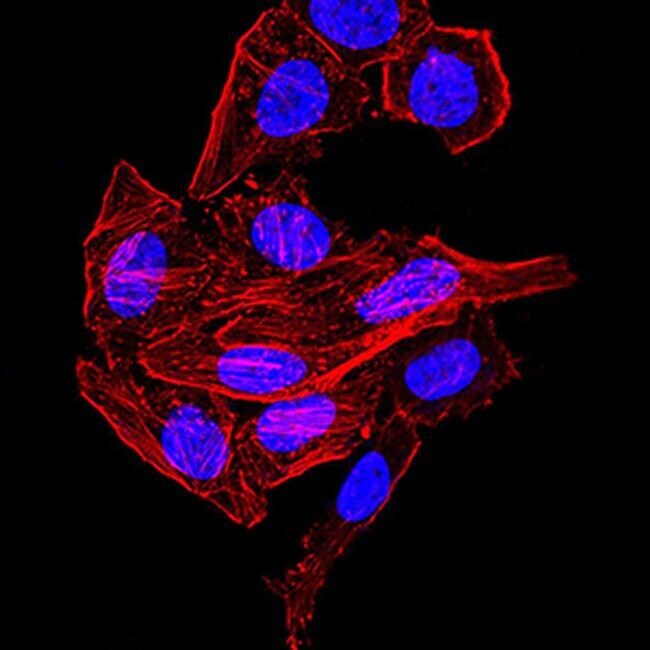
- Immunocytochemistry analysis of Synapsin 1 in GC-7901 cells (green). Sample was incubated with Synapsin 1 monoclonal antibody (Product # MA5-31919) using a dilution of 1:200-1:1000 followed by DRAQ5 fluorescent DNA dye (blue), and Alexa Fluor- 555 phalloidin (red labeled actin filaments) .
- Submitted by
- Invitrogen Antibodies (provider)
- Main image
- Experimental details
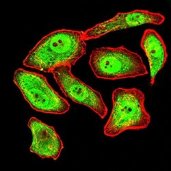
- Immunocytochemistry analysis of Synapsin 1 in HepG2 cells (green). Sample was incubated with Synapsin 1 monoclonal antibody (Product # MA5-31919) using a dilution of 1:200-1:1000 followed by DRAQ5 fluorescent DNA dye (blue), and Alexa Fluor- 555 phalloidin (red labeled actin filaments) .

- Submitted by
- Invitrogen Antibodies (provider)
- Main image
- Experimental details
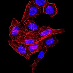
- Immunocytochemistry analysis of Synapsin 1 in GC-7901 cells. Sample was incubated with Synapsin 1 monoclonal antibody (Product # MA5-31919) using a dilution of 1:200-1:1000 followed by DRAQ5 fluorescent DNA dye (blue), and Alexa Fluor- 555 phalloidin (red labeled actin filaments) .

- Submitted by
- Invitrogen Antibodies (provider)
- Main image
- Experimental details
- Immunocytochemistry analysis of Synapsin 1 in GC-7901 cells (green). Sample was incubated with Synapsin 1 monoclonal antibody (Product # MA5-31919) using a dilution of 1:200-1:1000 followed by DRAQ5 fluorescent DNA dye (blue), and Alexa Fluor- 555 phalloidin (red labeled actin filaments) .

- Submitted by
- Invitrogen Antibodies (provider)
- Main image
- Experimental details
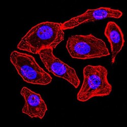
- Immunocytochemistry analysis of Synapsin 1 in HepG2 cells. Sample was incubated with Synapsin 1 monoclonal antibody (Product # MA5-31919) using a dilution of 1:200-1:1000 followed by DRAQ5 fluorescent DNA dye (blue), and Alexa Fluor- 555 phalloidin (red labeled actin filaments) .
- Submitted by
- Invitrogen Antibodies (provider)
- Main image

- Experimental details
- Immunocytochemistry analysis of Synapsin 1 in HepG2 cells (green). Sample was incubated with Synapsin 1 monoclonal antibody (Product # MA5-31919) using a dilution of 1:200-1:1000 followed by DRAQ5 fluorescent DNA dye (blue), and Alexa Fluor- 555 phalloidin (red labeled actin filaments) .

Supportive validation
- Submitted by
- Invitrogen Antibodies (provider)
- Main image

- Experimental details
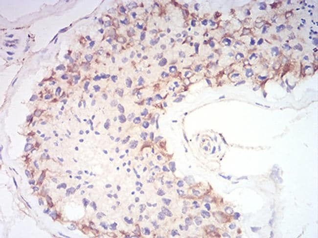
- Immunohistochemistry analysis of Synapsin 1 in paraffin-embedded testis tissue. Sample was incubated with Synapsin 1 monoclonal antibody (Product # MA5-31919) using a dilution of 1:200-1:1000 followed by DAB staining.
Supportive validation
- Submitted by
- Invitrogen Antibodies (provider)
- Main image

- Experimental details
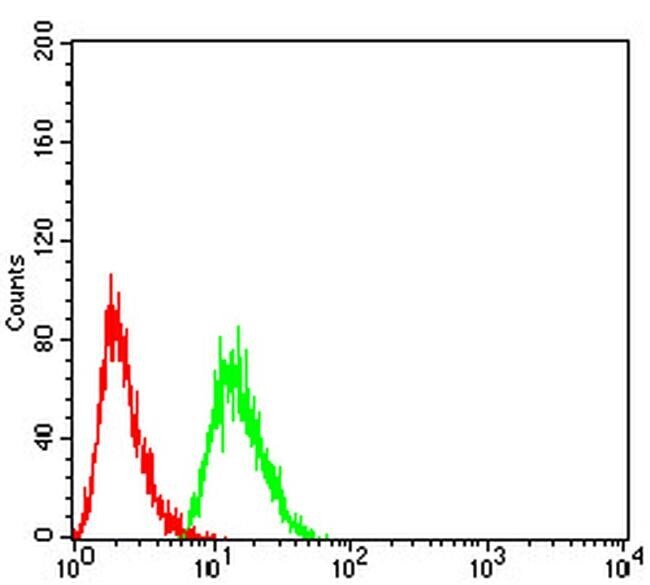
- Flow cytometry of Synapsin 1 in HeLa cells (green). Sample was incubated with Synapsin 1 monoclonal antibody (Product # MA5-31919) using a dilution of 1:200-1:400 followed by negative control (red).

- Submitted by
- Invitrogen Antibodies (provider)
- Main image

- Experimental details
- Flow cytometry of Synapsin 1 in HeLa cells (green). Sample was incubated with Synapsin 1 monoclonal antibody (Product # MA5-31919) using a dilution of 1:200-1:400 followed by negative control (red).