 Explore
 Validate
 Learn
Learn Western blot
Antibody data
- Antibody Data
- Antigen structure
- References [1]
- Comments [0]
- Validations
- Western blot [3]
Submit
Validation data
Reference
Comment
Report error
- Product number
- PA1-4673 - Provider product page

- Provider
- Invitrogen Antibodies
- Product name
- Synapsin 1 Polyclonal Antibody
- Antibody type
- Polyclonal
- Antigen
- Purifed from natural sources
- Description
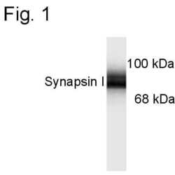
- This antibody is specific for the ~78 kDa Synapsin I protein doublet. Immunolabeling is blocked by preadsorption of the antibody with the protein used to generate the antibody.
Submitted references F1F0 ATP Synthase-Cyclophilin D Interaction Contributes to Diabetes-Induced Synaptic Dysfunction and Cognitive Decline.
Yan S, Du F, Wu L, Zhang Z, Zhong C, Yu Q, Wang Y, Lue LF, Walker DG, Douglas JT, Yan SS
Diabetes 2016 Nov;65(11):3482-3494
Diabetes 2016 Nov;65(11):3482-3494
No comments: Submit comment
Supportive validation
- Submitted by
- Invitrogen Antibodies (provider)
- Main image
- Experimental details
- Western blots of 10 µg of rat brain lysate showing specific immunolabeling of the ~78 kDa Synapsin I doublet.

- Submitted by
- Invitrogen Antibodies (provider)
- Main image

- Experimental details
- Western blot of Synapsin 1 in rat hippocampal lysate (10 µg) showing specific immunolabeling of a band at ~78 kDa corresponding to Synapsin 1 polyclonal antibody (Product # PA1-4673).

- Submitted by
- Invitrogen Antibodies (provider)
- Main image
- Experimental details
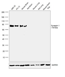
- Western blot was performed using anti-Synapsin1 Polyclonal Antibody (Product # PA1-4673) and a 78 kDa band was observed corresponding to Synapsin1 was observed across cell lines and tissues tested except Mouse and Rat Lung, Mouse Heart and Raji which are reported to be negative. Whole cell extracts of (30ug lysate) of IMR-32 (Lane 1), PC-12 (Lane 2), Raji (Lane 8), tissue extracts of Mouse Brain (Lane 3), Rat Brain (Lane 4), Mouse Lung (Lane 5), Rat Lung (Lane 6) and Mouse Heart (Lane 7) were electrophoresed using Novex® NuPAGE® 4-12 % Bis-Tris gel (Product # NP0322BOX). Resolved proteins were then transferred onto a nitrocellulose membrane (Product # IB23001) by iBlot® 2 Dry Blotting System (Product # IB21001).The blot was probed with the primary antibody (1:1000 dilution) and detected by chemiluminescence with Goat anti-Rabbit IgG (H+L) Superclonal™ Recombinant Secondary Antibody, HRP (Product # A27036, 1:4000 dilution). Chemiluminescent detection was performed using Novex® ECL Chemiluminescent Substrate Reagent Kit (Product # WP20005).
