Explore
 Validate
 Learn
Learn Western blot
 Immunocytochemistry
Antibody data
- Antibody Data
- Antigen structure
- References [3]
- Comments [0]
- Validations
- Immunocytochemistry [6]
- Immunohistochemistry [9]
- Other assay [1]
Submit
Validation data
Reference
Comment
Report error
- Product number
- PA5-85730 - Provider product page

- Provider
- Invitrogen Antibodies
- Product name
- BDNF Polyclonal Antibody
- Antibody type
- Polyclonal
- Antigen
- Synthetic peptide
- Description
- Keep as concentrated solution. Predicted reactivity: Mouse (100%), Rat (100%), Dog (100%), Cat (100%), Pig (100%), Chimpanzee (100%), Bovine (100%). Positive Control: mouse brain, rat brain, BDNF-transfected 293T. Store product as a concentrated solution. Centrifuge briefly prior to opening the vial.
- Reactivity
- Human, Mouse, Rat
- Host
- Rabbit
- Isotype
- IgG
- Vial size
- 100 μL
- Concentration
- 0.5 mg/mL
- Storage
- Store at 4°C short term. For long term storage, store at -20°C, avoiding freeze/thaw cycles.
Submitted references Neuregulin-1/ErbB4 signaling modulates Plasmodium falciparum HRP2-induced damage to brain cortical organoids.
Therapeutic Effect of Erythropoietin on Alzheimer's Disease by Activating the Serotonin Pathway.
Human Pluripotent Stem Cell-Derived Neural Progenitor Cells Promote Retinal Ganglion Cell Survival and Axon Recovery in an Optic Nerve Compression Animal Model.
Harbuzariu A, Nti A, Harp KO, Cespedes JC, Driss A, Stiles JK
iScience 2022 Jun 17;25(6):104407
iScience 2022 Jun 17;25(6):104407
Therapeutic Effect of Erythropoietin on Alzheimer's Disease by Activating the Serotonin Pathway.
Shim KH, Ha S, Choung JS, Choi JI, Kim DY, Kim JM, Kim M
International journal of molecular sciences 2022 Jul 24;23(15)
International journal of molecular sciences 2022 Jul 24;23(15)
Human Pluripotent Stem Cell-Derived Neural Progenitor Cells Promote Retinal Ganglion Cell Survival and Axon Recovery in an Optic Nerve Compression Animal Model.
Park M, Kim HM, Shin HA, Lee SH, Hwang DY, Lew H
International journal of molecular sciences 2021 Nov 20;22(22)
International journal of molecular sciences 2021 Nov 20;22(22)
No comments: Submit comment
Supportive validation
- Submitted by
- Invitrogen Antibodies (provider)
- Main image
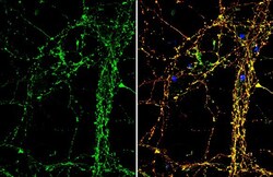
- Experimental details
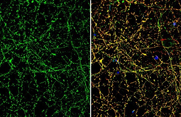
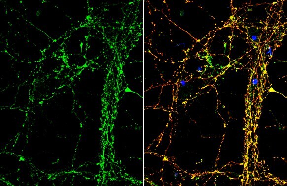
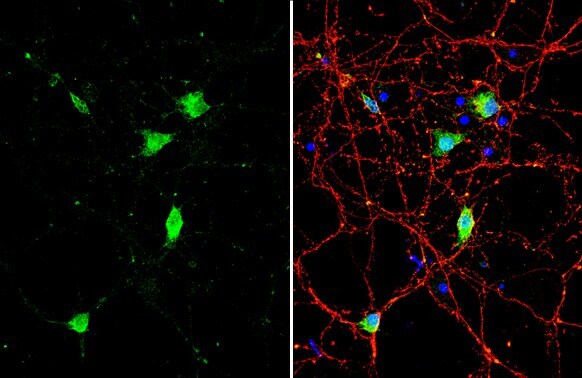
- Immunocytochemistry-Immunofluorescence analysis of BDNF was performed in DIV9 rat E18 primary cortical neuron cells fixed in 4% paraformaldehyde at RT for 15 min. Green: BDNF Polyclonal Antibody (Product # PA5-85730) diluted at 1:500. Red: Tau, stained by Tau antibody. Blue: Fluoroshield with DAPI.
- Submitted by
- Invitrogen Antibodies (provider)
- Main image
- Experimental details
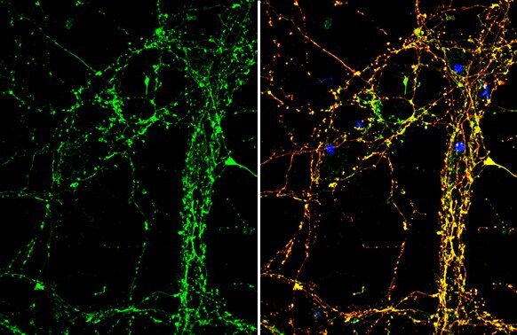
- Immunocytochemistry-Immunofluorescence analysis of BDNF was performed in DIV9 rat E18 primary cortical neuron cells fixed in 4% paraformaldehyde at RT for 15 min. Green: BDNF Polyclonal Antibody (Product # PA5-85730) diluted at 1:500. Red: Tau, stained by Tau antibody. Blue: Fluoroshield with DAPI.
- Submitted by
- Invitrogen Antibodies (provider)
- Main image
- Experimental details
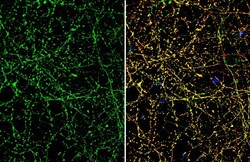
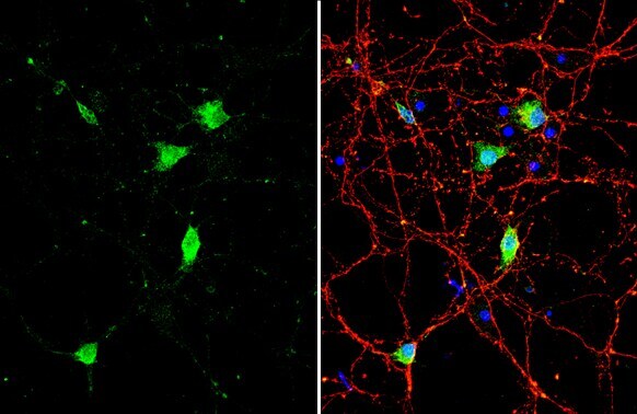
- BDNF Polyclonal Antibody detects BDNF protein by immunofluorescent analysis. Sample: DIV10 rat E18 primary cortical neuron cells were fixed in 4% paraformaldehyde at RT for 15 min. Green: BDNF stained by BDNF Polyclonal Antibody (Product # PA5-85730) diluted at 1:500. Red: Tau, stained by Phospho-Tau (Ser262) Polyclonal Antibody [GT287]diluted at 1:500. Blue: Fluoroshield with DAPI .
- Submitted by
- Invitrogen Antibodies (provider)
- Main image
- Experimental details
- Immunocytochemistry-Immunofluorescence analysis of BDNF was performed in DIV9 rat E18 primary cortical neuron cells fixed in 4% paraformaldehyde at RT for 15 min. Green: BDNF Polyclonal Antibody (Product # PA5-85730) diluted at 1:500. Red: Tau, stained by Tau antibody. Blue: Fluoroshield with DAPI.
- Submitted by
- Invitrogen Antibodies (provider)
- Main image
- Experimental details
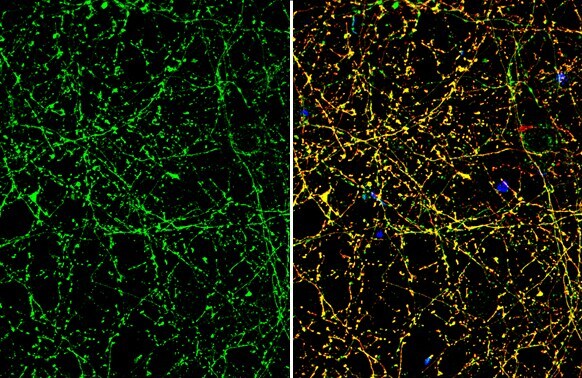
- Immunocytochemistry-Immunofluorescence analysis of BDNF was performed in DIV9 rat E18 primary cortical neuron cells fixed in 4% paraformaldehyde at RT for 15 min. Green: BDNF Polyclonal Antibody (Product # PA5-85730) diluted at 1:500. Red: Tau, stained by Tau antibody. Blue: Fluoroshield with DAPI.
- Submitted by
- Invitrogen Antibodies (provider)
- Main image
- Experimental details
- BDNF Polyclonal Antibody detects BDNF protein by immunofluorescent analysis. Sample: DIV10 rat E18 primary cortical neuron cells were fixed in 4% paraformaldehyde at RT for 15 min. Green: BDNF stained by BDNF Polyclonal Antibody (Product # PA5-85730) diluted at 1:500. Red: Tau, stained by Phospho-Tau (Ser262) Polyclonal Antibody [GT287]diluted at 1:500. Blue: Fluoroshield with DAPI .
Supportive validation
- Submitted by
- Invitrogen Antibodies (provider)
- Main image
- Experimental details
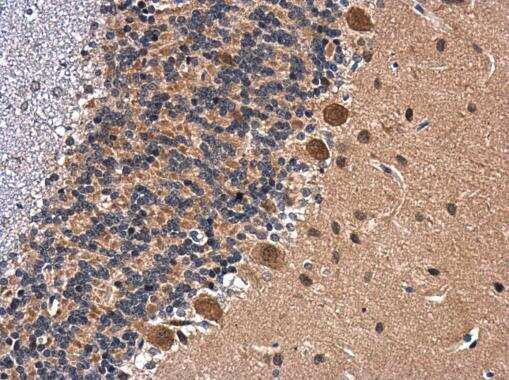
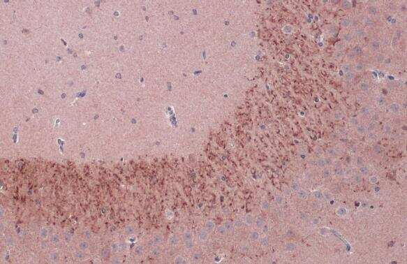
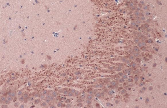
- BDNF Polyclonal Antibody detects BDNF protein at cytoplasm in rat brain by immunohistochemical analysis. Sample: Paraffin-embedded rat brain. BDNF Polyclonal Antibody (Product # PA5-85730) diluted at 1:500. Antigen Retrieval: Citrate buffer, pH 6.0, 15 min.
- Submitted by
- Invitrogen Antibodies (provider)
- Main image
- Experimental details
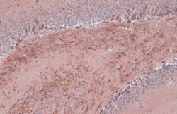
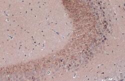
- Immunohistochemistry (Paraffin) analysis of BDNF was performed in paraffin-embedded rat hippocampus tissue using BDNF Polyclonal Antibody (Product # PA5-85730) at a dilution of 1:500. Antigen Retrieval: Citrate buffer, pH 6.0, 15 min.
- Submitted by
- Invitrogen Antibodies (provider)
- Main image
- Experimental details
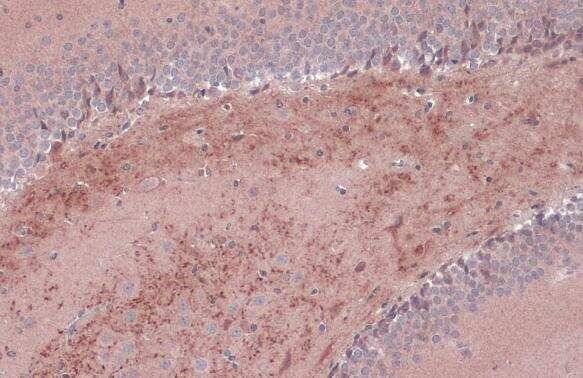
- Immunohistochemistry (Paraffin) analysis of BDNF was performed in paraffin-embedded rat hippocampus tissue using BDNF Polyclonal Antibody (Product # PA5-85730) at a dilution of 1:500. Antigen Retrieval: Citrate buffer, pH 6.0, 15 min.
- Submitted by
- Invitrogen Antibodies (provider)
- Main image
- Experimental details
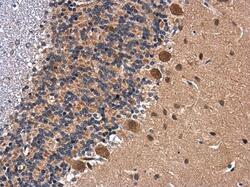
- Immunohistochemistry analysis of BDNF in paraffin-embedded rat brain using BDNF polyclonal antibody (Product # PA5-85730) at a dilution of 1:500.
- Submitted by
- Invitrogen Antibodies (provider)
- Main image
- Experimental details
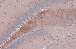
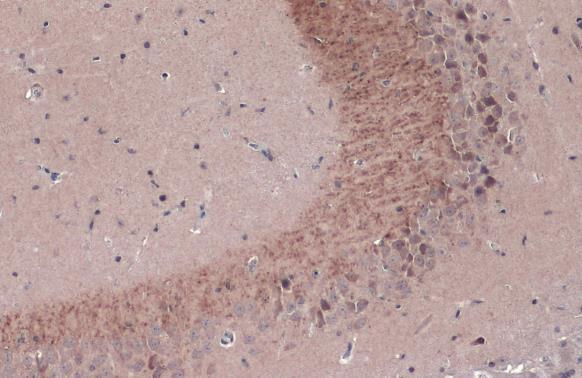
- BDNF Polyclonal Antibody detects BDNF protein at cytoplasm by immunohistochemical analysis. Sample: Paraffin-embedded mouse hippocampus. BDNF stained by BDNF Polyclonal Antibody (Product # PA5-85730) diluted at 1:1,000. Antigen Retrieval: Citrate buffer, pH 6.0, 15 min.
- Submitted by
- Invitrogen Antibodies (provider)
- Main image
- Experimental details
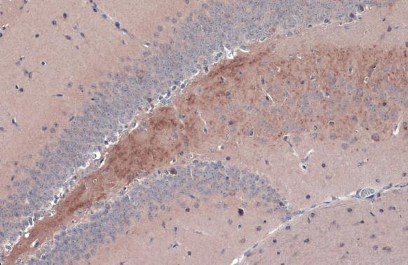
- BDNF Polyclonal Antibody detects BDNF protein at cytoplasm by immunohistochemical analysis. Sample: Paraffin-embedded mouse hippocampus. BDNF stained by BDNF Polyclonal Antibody (Product # PA5-85730) diluted at 1:1,000. Antigen Retrieval: Citrate buffer, pH 6.0, 15 min.
- Submitted by
- Invitrogen Antibodies (provider)
- Main image
- Experimental details
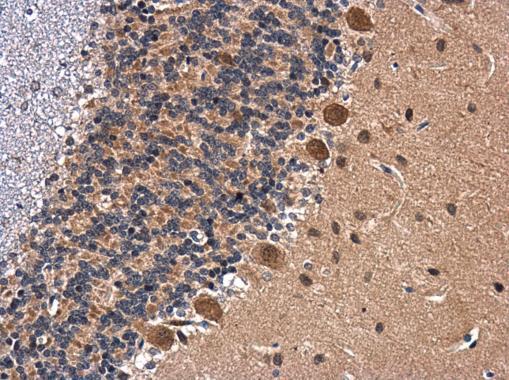
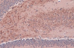
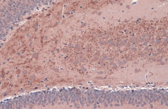
- BDNF Polyclonal Antibody detects BDNF protein at cytoplasm by immunohistochemical analysis. Sample: Paraffin-embedded rat brain. BDNF stained by BDNF Polyclonal Antibody (Product # PA5-85730) diluted at 1:1,000. Antigen Retrieval: Citrate buffer, pH 6.0, 15 min.
- Submitted by
- Invitrogen Antibodies (provider)
- Main image
- Experimental details
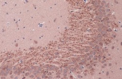
- BDNF Polyclonal Antibody detects BDNF protein at cytoplasm by immunohistochemical analysis. Sample: Paraffin-embedded rat brain. BDNF stained by BDNF Polyclonal Antibody (Product # PA5-85730) diluted at 1:1,000. Antigen Retrieval: Citrate buffer, pH 6.0, 15 min.
- Submitted by
- Invitrogen Antibodies (provider)
- Main image
- Experimental details
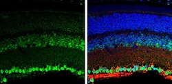
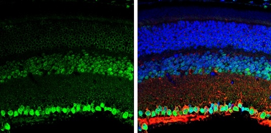
- Immunohistochemistry analysis of BDNF in paraffin-embedded adult mouse retina using BDNF polyclonal antibody (Product # PA5-85730) at a dilution of 1:250. Sample was then incubated with beta Tubulin 3/ TUJ1 and DAPI secondary antibody at a dilution of 1:500.
Supportive validation
- Submitted by
- Invitrogen Antibodies (provider)
- Main image
- Experimental details
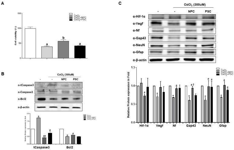
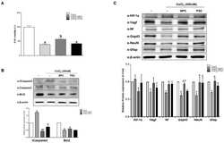
- Figure 2 Human NPCs have the function of recovering damaged R28 cells. R28 cells were cultured with NPCs or hPSCs 3 h prior to CoCl 2 treatment. Then, the R28 cells were treated with CoCl 2 (300 muM). ( A ) Viability assays performed after 24 h. Data are expressed as percentage (mean +- SEM) of viable cells compared to those in the control group. Significantly different values between groups are indicated by different letters ( p < 0.05). ( B ) Apoptosis-related protein expressions were also determined. ( C ) Western blot analyses of target protein expression levels, using R28 lysates with CoCl 2 . The quantified values of target protein expression are presented (bottom panel) (* p < 0.05 vs. the control; # p < 0.05, ## p < 0.01 vs. CoCl 2; + p < 0.05 vs. PSCs).