Explore
 Validate
 Learn
Learn Western blot
 Immunocytochemistry
Antibody data
- Antibody Data
- Antigen structure
- References [1]
- Comments [0]
- Validations
- Immunocytochemistry [3]
- Immunohistochemistry [7]
- Flow cytometry [1]
Submit
Validation data
Reference
Comment
Report error
- Product number
- MA5-32783 - Provider product page

- Provider
- Invitrogen Antibodies
- Product name
- GCLM Recombinant Rabbit Monoclonal Antibody (JM39-61)
- Antibody type
- Monoclonal
- Antigen
- Recombinant full-length protein
- Description
- Recombinant rabbit monoclonal antibodies are produced using in vitro expression systems. The expression systems are developed by cloning in the specific antibody DNA sequences from immunoreactive rabbits. Then, individual clones are screened to select the best candidates for production. The advantages of using recombinant rabbit monoclonal antibodies include: better specificity and sensitivity, lot-to-lot consistency, animal origin-free formulations, and broader immunoreactivity to diverse targets due to larger rabbit immune repertoire.
- Reactivity
- Human, Mouse, Rat
- Host
- Rabbit
- Isotype
- IgG
- Antibody clone number
- JM39-61
- Vial size
- 100 μL
- Concentration
- 1 mg/mL
- Storage
- Store at 4°C short term. For long term storage, store at -20°C, avoiding freeze/thaw cycles.
Submitted references Elevated type I interferon responses potentiate metabolic dysfunction, inflammation, and accelerated aging in mtDNA mutator mice.
Lei Y, Guerra Martinez C, Torres-Odio S, Bell SL, Birdwell CE, Bryant JD, Tong CW, Watson RO, West LC, West AP
Science advances 2021 May;7(22)
Science advances 2021 May;7(22)
No comments: Submit comment
Supportive validation
- Submitted by
- Invitrogen Antibodies (provider)
- Main image

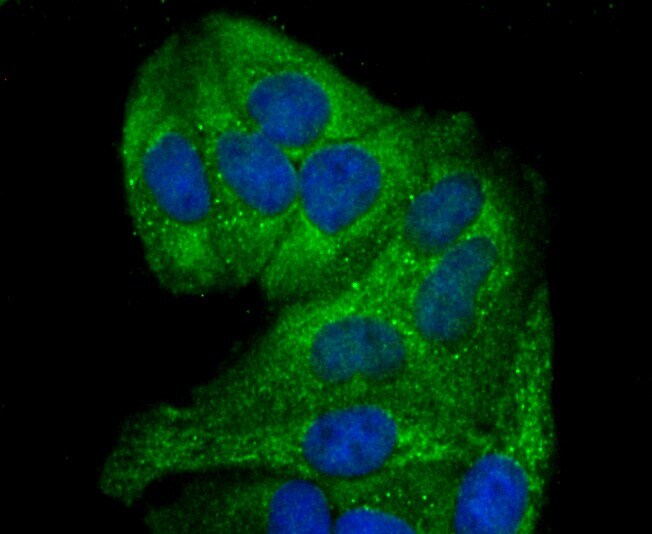
- Experimental details
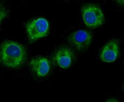
- Immunocytochemical analysis of GCLM in Hela cells using a GCLM Monoclonal antibody (Product # MA5-32783) as seen in green. The nuclear counter stain is DAPI (blue). Cells were fixed in paraformaldehyde, permeabilised with 0.25% Triton X100/PBS.
- Submitted by
- Invitrogen Antibodies (provider)
- Main image
- Experimental details
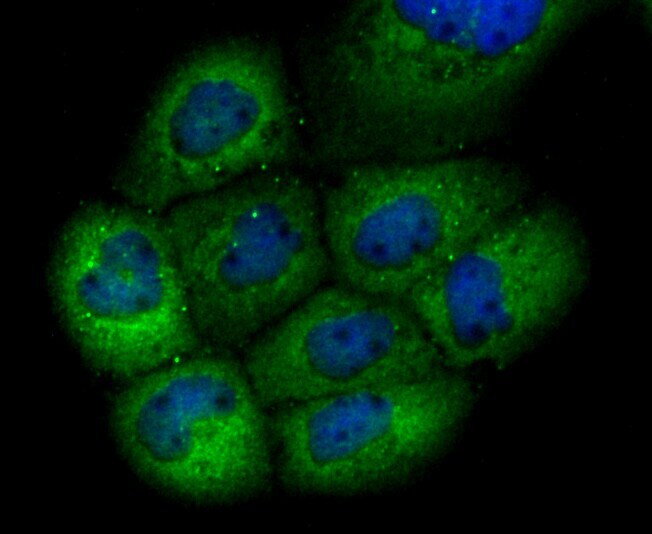
- Immunocytochemistry analysis of GCLM with Hela cells (green). Formalin fixed cells were permeabilized with 0.1% Triton X-100 in TBS (10 min, room temp) and blocked with 1% Blocker BSA (15 min, room temp), incubated with recombinant monoclonal GCLM (Product # MA5-32783) at a dilution of 1:50 for 1 hour at room temperature, washed with PBS, followed by Alexa Fluor 488 Goat anti-Rabbit IgG at 1:1,000 dilution. DAPI was used to stain the cell nuclei (blue).
- Submitted by
- Invitrogen Antibodies (provider)
- Main image
- Experimental details
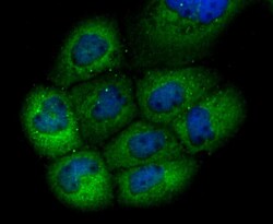
- Immunocytochemistry analysis of GCLM with A549 cells (green). Formalin fixed cells were permeabilized with 0.1% Triton X-100 in TBS (10 min, room temp) and blocked with 1% Blocker BSA (15 min, room temp), incubated with recombinant monoclonal GCLM (Product # MA5-32783) at a dilution of 1:50 for 1 hour at room temperature, washed with PBS, followed by Alexa Fluor 488 Goat anti-Rabbit IgG at 1:1,000 dilution. DAPI was used to stain the cell nuclei (blue).
Supportive validation
- Submitted by
- Invitrogen Antibodies (provider)
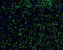
- Main image

- Experimental details
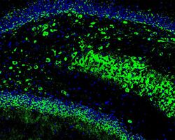
- Immunohistochemistry analysis of GCLM with frozen mouse hippocampus. The tissues were blocked in 3% BSA (30 min, room temp), washed with PBS, incubated with recombinant monoclonal GCLM (Product # MA5-32783) at a dilution of 1:50 dilution (overnight, 4°C, washed with PBS), followed by Alexa Fluor Goat Anti-Rabbit IgG H&L 488 at 1:200 dilution. DAPI was used to stain the cell nuclei (blue).

- Submitted by
- Invitrogen Antibodies (provider)
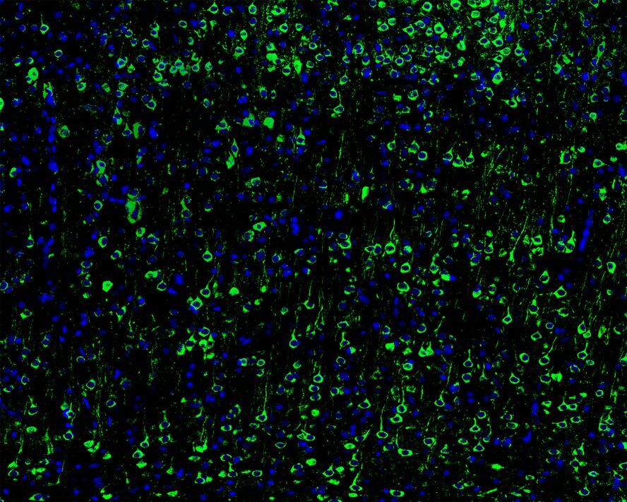
- Main image
- Experimental details
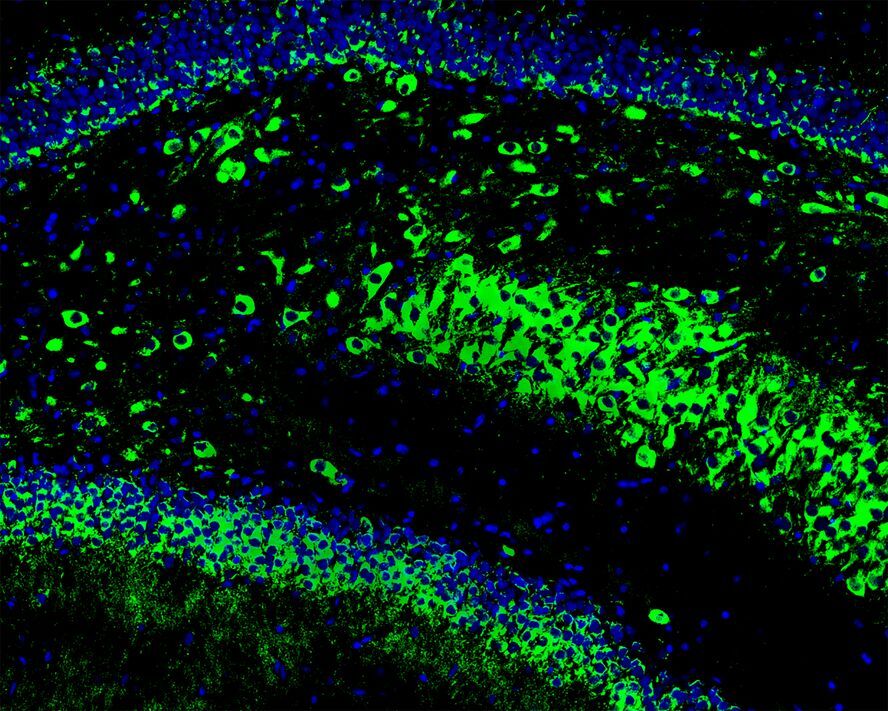
- Immunohistochemistry analysis of GCLM with frozen mouse cerebral cortex. The tissues were blocked in 3% BSA (30 min, room temp), washed with PBS, incubated with recombinant monoclonal GCLM (Product # MA5-32783) at a dilution of 1:100 dilution (overnight, 4°C, washed with PBS), followed by Alexa Fluor Goat Anti-Rabbit IgG H&L 488 at 1:200 dilution. DAPI was used to stain the cell nuclei (blue).

- Submitted by
- Invitrogen Antibodies (provider)
- Main image

- Experimental details
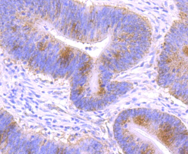
- Immunohistochemical analysis of GCLM of paraffin-embedded Human colon cancer tissue using a GCLM Monoclonal antibody (Product #MA5-32783). Counter stained with hematoxylin.
- Submitted by
- Invitrogen Antibodies (provider)
- Main image
- Experimental details
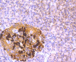
- Immunohistochemical analysis of GCLM of paraffin-embedded Human pancreas tissue using a GCLM Monoclonal antibody (Product #MA5-32783). Counter stained with hematoxylin.

- Submitted by
- Invitrogen Antibodies (provider)
- Main image
- Experimental details
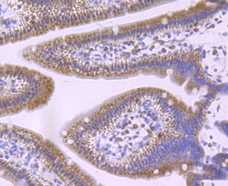
- Immunohistochemical analysis of GCLM of paraffin-embedded Mouse small intestine tissue using a GCLM Monoclonal antibody (Product #MA5-32783). Counter stained with hematoxylin.

- Submitted by
- Invitrogen Antibodies (provider)
- Main image
- Experimental details
- Immunohistochemistry analysis of GCLM with frozen mouse hippocampus. The tissues were blocked in 3% BSA (30 min, room temp), washed with PBS, incubated with recombinant monoclonal GCLM (Product # MA5-32783) at a dilution of 1:50 dilution (overnight, 4°C, washed with PBS), followed by Alexa Fluor Goat Anti-Rabbit IgG H&L 488 at 1:200 dilution. DAPI was used to stain the cell nuclei (blue).
- Submitted by
- Invitrogen Antibodies (provider)
- Main image
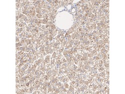
- Experimental details
- Immunohistochemistry analysis of paraffin-embedded GCLM in rat liver tissue. The section was pre-treated using heat mediated antigen retrieval with Tris-EDTA buffer (pH 9.0) for 20 minutes. The tissues were blocked in 1% BSA for 20 minutes at room temperature, washed with ddH2O and PBS. Incubation was done with monoclonal GCLM antibody (Product # MA5-32783) at a dilution of 1:200 (1h at RT). The detection was performed using an HRP conjugated compact polymer system. DAB was used as the chromogen. Tissues were counterstained with hematoxylin and mounted with DPX.

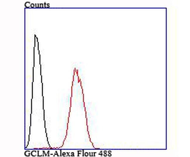
Supportive validation
- Submitted by
- Invitrogen Antibodies (provider)
- Main image
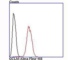
- Experimental details
- Flow Cytometric analysis of GCLM in Hela cells using a GCLM Monoclonal Antibody (Product # MA5-32783) at a dilution of 1:100, as seen in red compared with an unlabelled control (cells without incubation with primary antibody; black).