 Explore
 Validate
 Learn
Learn Immunocytochemistry
Antibody data
- Antibody Data
- Antigen structure
- References [1]
- Comments [0]
- Validations
- Immunocytochemistry [1]
- Flow cytometry [2]
Submit
Validation data
Reference
Comment
Report error
- Product number
- MAB5051 - Provider product page

- Provider
- R&D Systems
- Product name
- Human BMPR-IB/ALK-6 Antibody
- Antibody type
- Monoclonal
- Description
- Protein A or G purified from hybridoma culture supernatant. Detects human BMPR-IB/ALK-6 in direct ELISAs. In direct ELISAs, no cross-reactivity with recombinant mouse BMPR-IB is observed.
- Reactivity
- Human
- Host
- Mouse
- Conjugate
- Unconjugated
- Antigen sequence
O00238- Isotype
- IgG
- Antibody clone number
- 477914
- Vial size
- 100 ug
- Concentration
- LYOPH
- Storage
- Use a manual defrost freezer and avoid repeated freeze-thaw cycles. 12 months from date of receipt, -20 to -70 °C as supplied. 1 month, 2 to 8 °C under sterile conditions after reconstitution. 6 months, -20 to -70 °C under sterile conditions after reconstitution.
Submitted references Endocytosis contributes to BMP2-induced Smad signalling and neuronal growth.
Hegarty SV, Sullivan AM, O'Keeffe GW
Neuroscience letters 2017 Mar 16;643:32-37
Neuroscience letters 2017 Mar 16;643:32-37
No comments: Submit comment
Supportive validation
- Submitted by
- R&D Systems (provider)
- Main image

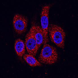
- Experimental details
- BMPR-IB/ALK-6 in PC-3 Human Cell Line. BMPR-IB/ALK-6 was detected in immersion fixed PC-3 human prostate cancer cell line using Mouse Anti-Human BMPR-IB/ALK-6 Monoclonal Antibody (Catalog # MAB5051) at 10 µg/mL for 3 hours at room temperature. Cells were stained using the NorthernLights™ 557-conjugated Anti-Mouse IgG Secondary Antibody (red; Catalog # NL007) and counterstained with DAPI (blue). Specific staining was localized to the cytoplasm and cell surface. View our protocol for Fluorescent ICC Staining of Cells on Coverslips.
Supportive validation
- Submitted by
- R&D Systems (provider)
- Main image
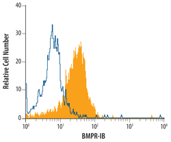
- Experimental details
- Detection of BMPR-IB/ALK-6 in PC-3 Human Cell Line by Flow Cytometry. PC-3 human prostate cancer cell line was stained with Mouse Anti-Human BMPR-IB/ ALK-6 Monoclonal Antibody (Catalog # MAB5051, filled histogram) or isotype control antibody (Catalog # MAB0041, open histogram), followed by Allophycocyanin-conjugated Anti-Mouse IgG F(ab')2 Secondary Antibody (Catalog # F0101B).

- Submitted by
- R&D Systems (provider)
- Main image
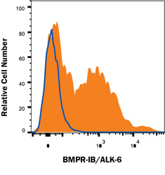
- Experimental details
- Detection of BMPR-IB/ALK-6 in Human iPS cells differentiated to Mesoderm by Flow Cytometry. Human iPS cells differentiated to mesoderm (using Catalog # SC030B) were stained with Mouse Anti-Human BMPR-IB/ALK-6 Monoclonal Antibody (Catalog # MAB5051, filled histogram) or isotype control antibody (Catalog # MAB0041, open histogram) followed by anti-Mouse IgG PE-conjugated secondary antibosy (Catalog # F0101B). View our protocol for Staining Membrane-associated Proteins.
