Explore
 Validate
 Learn
Learn Western blot
Antibody data
- Antibody Data
- Antigen structure
- References [0]
- Comments [0]
- Validations
- Western blot [1]
- Immunocytochemistry [1]
- Immunohistochemistry [1]
- Flow cytometry [1]
Submit
Validation data
Reference
Comment
Report error
- Product number
- TA328689 - Provider product page

- Provider
- OriGene
- Product name
- Rabbit Polyclonal Anti-Cannabinoid Receptor 2 (extracellular)
- Antibody type
- Polyclonal
- Description
- Rabbit Polyclonal Anti-Cannabinoid Receptor 2 (extracellular)
- Host
- Rabbit
- Conjugate
- Unconjugated
- Epitope
- CNR2
- Antibody clone number
- NULL
- Vial size
- 200 µl
- Concentration
- NULL
No comments: Submit comment
Supportive validation
- Submitted by
- OriGene (provider)
- Main image
- Experimental details
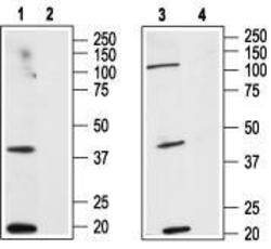
- Western blot analysis of HL-60 (human Promyelocytic leukemia) (lanes 1and 2) and MCF-7 (human adenocarcinoma, mammary gland) (lanes 3 and 4) cell line lysates: 1, 3. Anti-Cannabinoid Receptor 2 (extracellular) antibody, (1:200). 2, 4. Anti-Cannabinoid Receptor 2 (extracellular) antibody, preincubated with the control peptide antigen.
- Validation comment
- WB

Supportive validation
- Submitted by
- OriGene (provider)
- Main image
- Experimental details
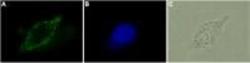
- Expression of CB2R in human LNCaP. Immunocytochemical staining of human prostate carcinoma (LNCaP). A. Cells were stained with Anti-Cannabinoid Receptor 2 (extracellular) antibody, (1:50), followed by goat-anti-rabbit-AlexaFluor-480 secondary antibody. B. Nuclear staining of LNCaP cells with Hoechst 33342. C. Live intact LNCaP cells.
- Validation comment
- IF
Supportive validation
- Submitted by
- OriGene (provider)
- Main image
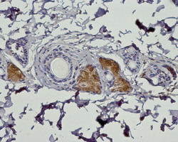
- Experimental details
- Expression of Cannabinoid Receptor 2 in rat dermis.Immunohistochemical staining of paraffin embedded sections of rat dermis using Anti-Cannabinoid Receptor 2 (extracellular) antibody (1:100). Cannabinoid Receptor 2 (brown) is detected in sebaceous glands in the reticular dermis. Hematoxilin was used as the Counterstain.
- Validation comment
- IHC

Supportive validation
- Submitted by
- OriGene (provider)
- Main image
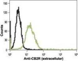
- Experimental details
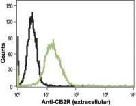
- Indirect flow cytomtery analysis in live intact human HL-60 promyelocytic leukemia cells: black line, Cells + goat-anti-rabbit-FITC alone. green line, Cells + Anti-Cannabinoid Receptor 2 (extracellular) antibody, (5 ug) + goat-anti-rabbit-FITC.
- Validation comment
- FC