Explore
 Validate
 Learn
Learn Western blot
 Immunocytochemistry
Antibody data
- Antibody Data
- Antigen structure
- References [1]
- Comments [0]
- Validations
- Immunocytochemistry [1]
- Immunohistochemistry [2]
- Other assay [2]
Submit
Validation data
Reference
Comment
Report error
- Product number
- PA5-20404 - Provider product page

- Provider
- Invitrogen Antibodies
- Product name
- STEAP1 Polyclonal Antibody
- Antibody type
- Polyclonal
- Antigen
- Synthetic peptide
- Description
- A suggested positive control is human spleen tissue lysate. PA5-20404 can be used with blocking peptide PEP-0521.
- Reactivity
- Human, Mouse, Rat
- Host
- Rabbit
- Isotype
- IgG
- Vial size
- 100 μg
- Concentration
- 1 mg/mL
- Storage
- Maintain refrigerated at 2-8°C for up to 3 months. For long term storage store at -20°C
Submitted references STEAP1 facilitates metastasis and epithelial-mesenchymal transition of lung adenocarcinoma via the JAK2/STAT3 signaling pathway.
Huo SF, Shang WL, Yu M, Ren XP, Wen HX, Chai CY, Sun L, Hui K, Liu LH, Wei SH, Wang XX, Wang Y, Tian YX
Bioscience reports 2020 Jun 26;40(6)
Bioscience reports 2020 Jun 26;40(6)
No comments: Submit comment
Supportive validation
- Submitted by
- Invitrogen Antibodies (provider)
- Main image
- Experimental details
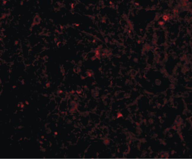
- Immunofluorescent analysis of human spleen tissue using a STEAP1 polyclonal antibody (Product # PA5-20404) at a 20 µg/mL dilution.
Supportive validation
- Submitted by
- Invitrogen Antibodies (provider)
- Main image

- Experimental details
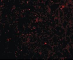
- Immunofluorescence of STEAP1 in human spleen tissue with STEAP1 Polyclonal Antibody (Product # PA5-20404) at 20 µg/mL.
- Submitted by
- Invitrogen Antibodies (provider)
- Main image
- Experimental details
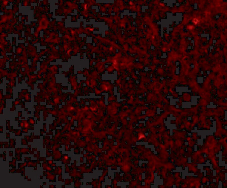
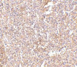
- Immunohistochemistry of STEAP1 in human spleen tissue with STEAP1 Polyclonal Antibody (Product # PA5-20404) at 2.5 µg/mL.

Supportive validation
- Submitted by
- Invitrogen Antibodies (provider)
- Main image

- Experimental details
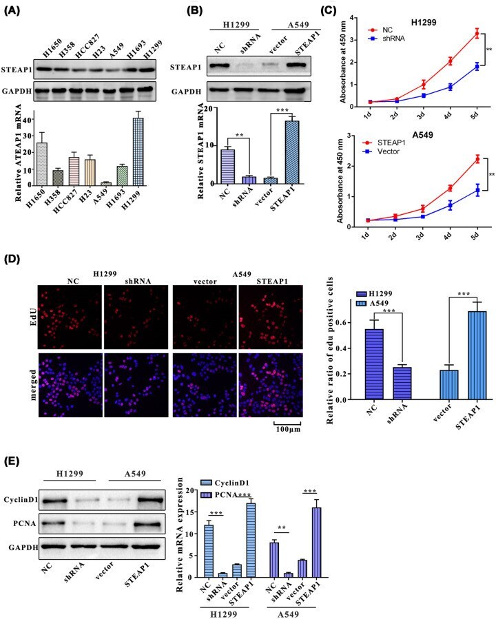
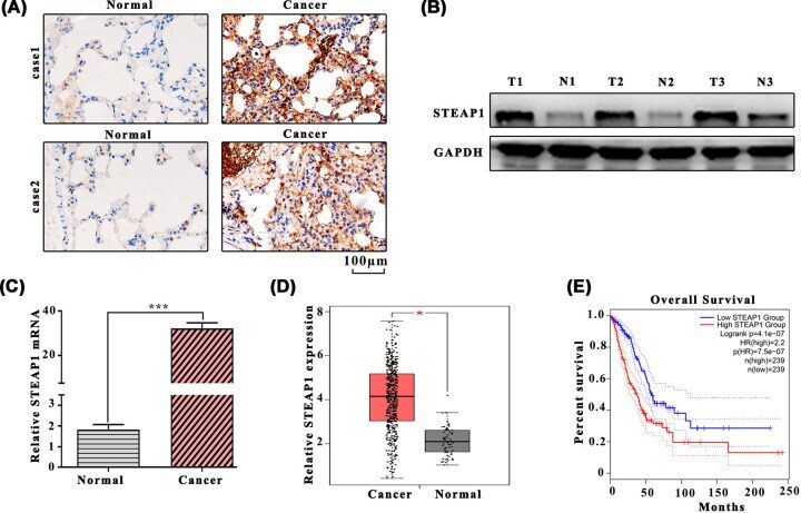
- Figure 1 The expression level of STEAP1 in LUAD tissues and para-carcinoma normal tissues ( A ) Western blot analysis of LUAD tissues and para-carcinoma normal tissues. ( B ) Western blot analysis of STEAP1 and GAPDH. ( C ) Detection of mRNA expression in LUAD tissues and para-carcinoma normal tissues by Q-PCR. ( D ) Relative STEAP1 expression among LUAD tissues and para-carcinoma normal tissues in the GEPIA dataset. ( E ) Survival percentage of the high and low STEAP1 group in the GEPIA dataset.
- Submitted by
- Invitrogen Antibodies (provider)
- Main image

- Experimental details
- Figure 2 STEAP1 promotes the proliferation of LUAD cells ( A ) Western blot and Q-PCR assay of H1650, H358, HCC827, H1299, H23, A549, and H1693. ( B ) Western blot and Q-PCR assay of H1299NC, H1299shRNA, A549vector, and A549STEAP1. ( C ) Cell proliferation in H1299NC, H1299shRNA, A549vector, and A549STEAP1 was measured using CCK8 reagent for five consecutive days. ( D ) Relative ratio of EdU positive cells of H1299NC, H1299shRNA, A549vector, and A549STEAP1. ( E ) Western blot and Q-PCR assay of PCNA and cyclinD1.