Explore
 Validate
 Learn
Learn Western blot
 ELISA
ELISAAntibody data
- Antibody Data
- Antigen structure
- References [1]
- Comments [0]
- Validations
- Western blot [1]
- Immunohistochemistry [2]
- Flow cytometry [1]
Submit
Validation data
Reference
Comment
Report error
- Product number
- NB300-953 - Provider product page

- Provider
- Novus Biologicals
- Proper citation
- Novus Cat#NB300-953, RRID:AB_10001859
- Product name
- Goat Polyclonal PARD6A Antibody
- Antibody type
- Polyclonal
- Description
- Immunogen affinity purified. This antibody is expected to recognise both reported isoforms (NP_058644.1
- Reactivity
- Human
- Host
- Goat
- Isotype
- IgG
- Vial size
- 0.1 mg
- Concentration
- 0.5 mg/ml
- Storage
- Store at -20C. Avoid freeze-thaw cycles.
Submitted references The mammalian homologue of the Caenorhabditis elegans polarity protein PAR-6 is a binding partner for the Rho GTPases Cdc42 and Rac1.
Johansson A, Driessens M, Aspenström P
Journal of cell science 2000 Sep;113 ( Pt 18):3267-75
Journal of cell science 2000 Sep;113 ( Pt 18):3267-75
No comments: Submit comment
Supportive validation
- Submitted by
- Novus Biologicals (provider)
- Main image
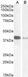
- Experimental details
- Western Blot: PARD6A Antibody [NB300-953] - (2ug/ml) staining of Jurkat (A) and U251 (B) cell lysate (35ug protein in RIPA buffer). Detected by chemiluminescence.
Supportive validation
- Submitted by
- Novus Biologicals (provider)
- Main image
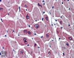
- Experimental details
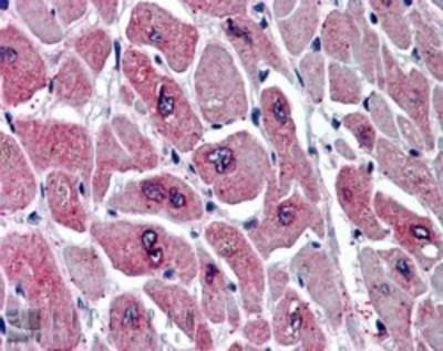
- Immunohistochemistry-Paraffin: PARD6A Antibody [NB300-953] - (5ug/ml) staining of paraffin embedded Human Heart. Steamed antigen retrieval with citrate buffer pH 6, AP-staining.
- Submitted by
- Novus Biologicals (provider)
- Main image
- Experimental details
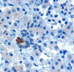
- Immunohistochemistry-Paraffin: PARD6A Antibody [NB300-953] - (10ug/ml) staining of paraffin embedded Human Human Pancreas. Microwaved antigen retrieval with Tris/EDTA buffer pH9, HRP-staining.

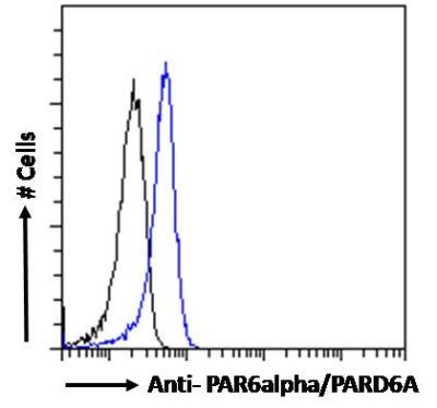
Supportive validation
- Submitted by
- Novus Biologicals (provider)
- Main image
- Experimental details
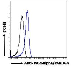
- Flow Cytometry: PARD6A Antibody [NB300-953] - Analysis of paraformaldehyde fixed Jurkat cells (blue line), permeabilized with 0.5% Triton. Primary incubation 1hr (10ug/ml) followed by Alexa Fluor 488 secondary antibody (1ug/ml). IgG control: Unimmunized goat IgG (black line) followed by Alexa Fluor 488 secondary antibody.