Explore
 Validate
 Learn
Learn Western blot
 Immunohistochemistry
Antibody data
- Antibody Data
- Antigen structure
- References [1]
- Comments [0]
- Validations
- Immunohistochemistry [5]
- Other assay [1]
Submit
Validation data
Reference
Comment
Report error
- Product number
- PA5-51995 - Provider product page

- Provider
- Invitrogen Antibodies
- Product name
- WIPF1 Polyclonal Antibody
- Antibody type
- Polyclonal
- Antigen
- Recombinant protein fragment
- Description
- Immunogen sequence: PPPPVSRNGS TSRALPATPQ LPSRSGVDSP RSGPRPPLPP DRPSAGAPPP PPPSTSIRNG FQDSPCEDEW ESRFYFHPIS DLPPPEPYVQ TTKSYPSKLA RNESRSGSNR RERGAP Highest antigen sequence identity to the following orthologs: Mouse - 92%, Rat - 90%.
- Reactivity
- Human
- Host
- Rabbit
- Isotype
- IgG
- Vial size
- 100 μL
- Concentration
- 0.05 mg/mL
- Storage
- Store at 4°C short term. For long term storage, store at -20°C, avoiding freeze/thaw cycles.
Submitted references FLI1 Induces Megakaryopoiesis Gene Expression Through WAS/WIP-Dependent and Independent Mechanisms; Implications for Wiskott-Aldrich Syndrome.
Wang C, Sample KM, Gajendran B, Kapranov P, Liu W, Hu A, Zacksenhaus E, Li Y, Hao X, Ben-David Y
Frontiers in immunology 2021;12:607836
Frontiers in immunology 2021;12:607836
No comments: Submit comment
Supportive validation
- Submitted by
- Invitrogen Antibodies (provider)
- Main image
- Experimental details
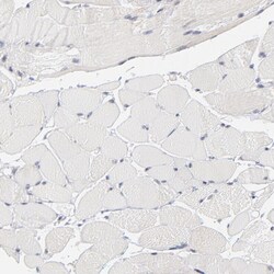
- Immunohistochemical analysis of WIPF1 in human skeletal muscle using WIPF1 Polyclonal Antibody (Product # PA5-51995) shows no cytoplasmic positivity in myocytes as expected.
- Submitted by
- Invitrogen Antibodies (provider)
- Main image
- Experimental details
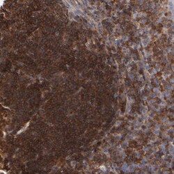
- Immunohistochemical analysis of WIPF1 in human tonsil using WIPF1 Polyclonal Antibody (Product # PA5-51995) shows strong cytoplasmic positivity in germinal center cells.

- Submitted by
- Invitrogen Antibodies (provider)
- Main image
- Experimental details
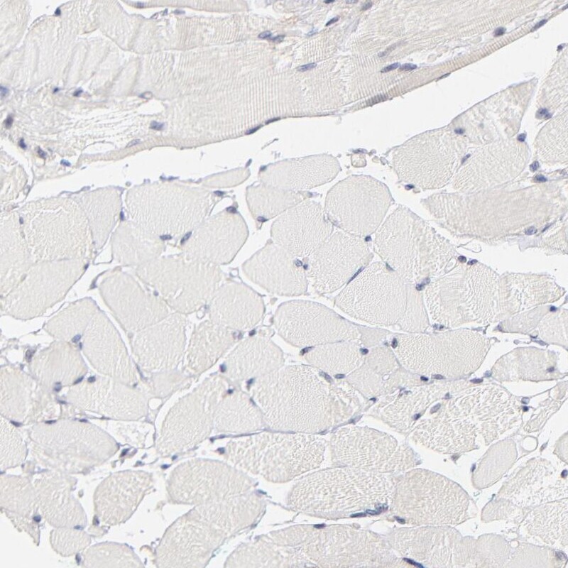
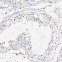
- Immunohistochemical analysis of WIPF1 in human testis using WIPF1 Polyclonal Antibody (Product # PA5-51995) shows no cytoplasmic positivity in cells in seminiferous ducts as expected.
- Submitted by
- Invitrogen Antibodies (provider)
- Main image
- Experimental details
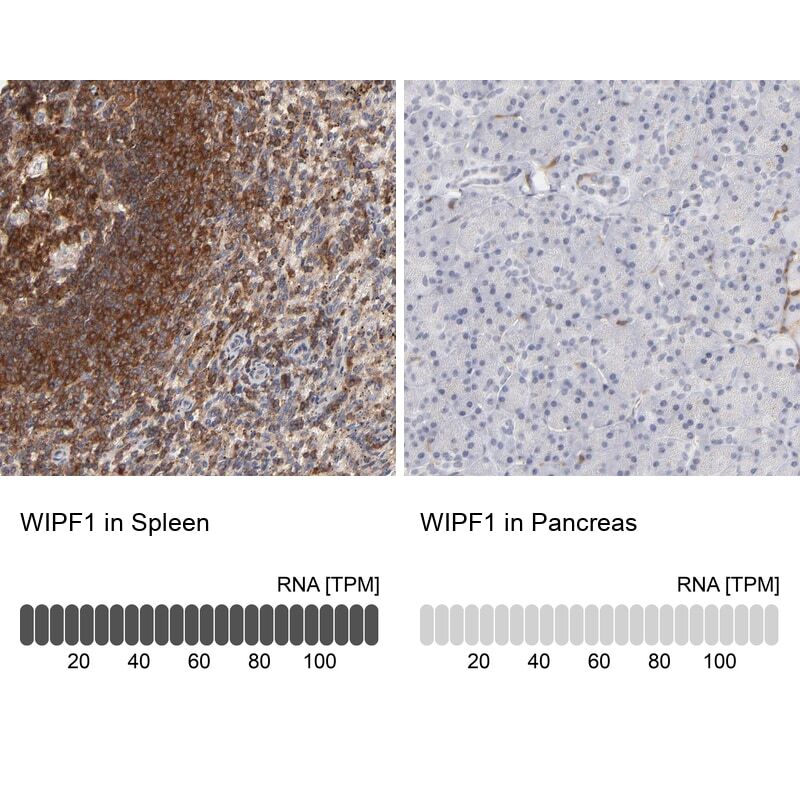
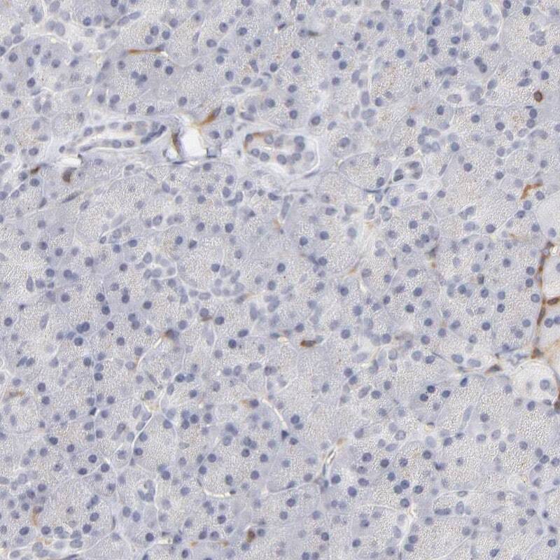
- Immunohistochemical staining of WIPF1 in human spleen and pancreas tissues using WIPF1 Polyclonal Antibody (Product # PA5-51995). Corresponding WIPF1 RNA-seq data are presented for the same tissues.
- Submitted by
- Invitrogen Antibodies (provider)
- Main image
- Experimental details
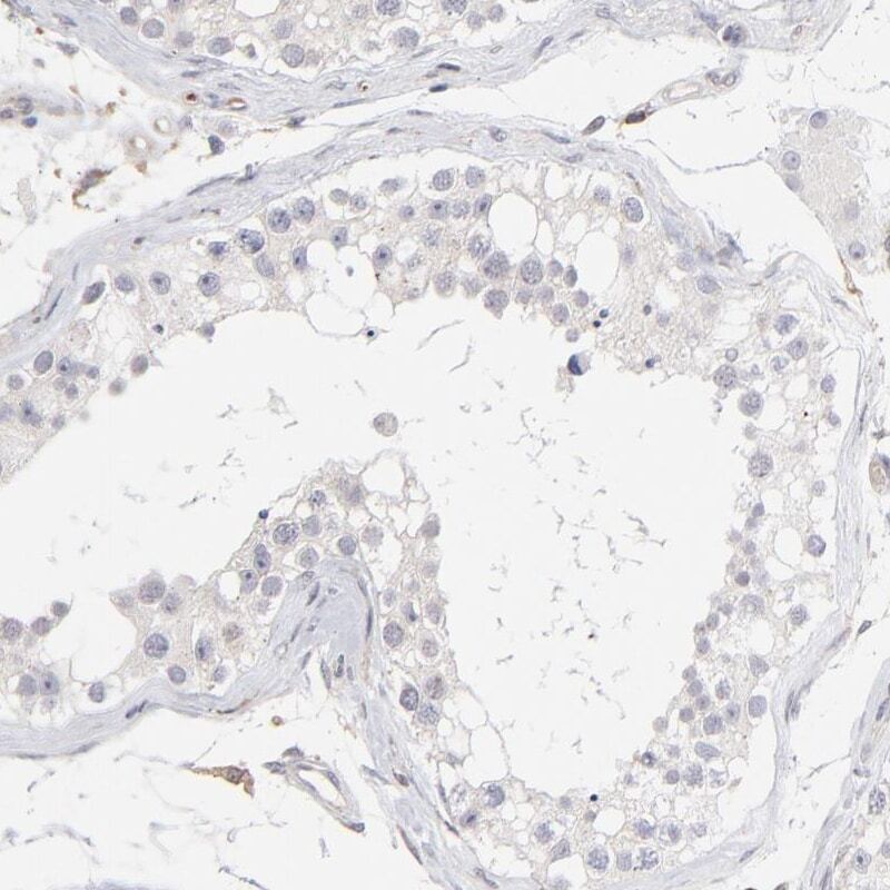
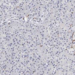
- Immunohistochemical staining of WIPF1 in human pancreas using WIPF1 Polyclonal Antibody (Product # PA5-51995) shows low expression as expected.
Supportive validation
- Submitted by
- Invitrogen Antibodies (provider)
- Main image
- Experimental details
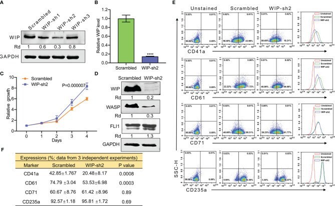
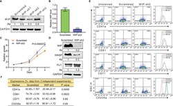
- Figure 5 ShRNA mediated knockdown of WIP suppresses the percentage of CD41a and CD61 expressing HEL cells. (A) Depletion of WIP in HEL cells using three shRNAs (shRNA1-shRNA3). (B) Knockdown of WIP in HEL cells using shRNA2 (WIP-sh2), as determined by Q-RT-PCR. (C) Knockdown of WIP in HEL cells (WIPF-sh2) accelerated proliferation when compared to scrambled-transfected HEL cells. (D) Western blot of WIP-sh2 and control cells for expression of WIP, FLI1 and WASP. (E) Flow cytometry analysis of WIP-sh2 and control scrambled cells for expression of indicated megakaryocytic and erythroid markers. The unstained identical plots (left most panels) were used as negative control. (F) Summary and statistics of flow cytometry experiments (n = 3). ****P