Explore
 Validate
 Learn
Learn Western blot
 Immunocytochemistry
Antibody data
- Antibody Data
- Antigen structure
- References [0]
- Comments [0]
- Validations
- Immunocytochemistry [1]
- Immunohistochemistry [1]
Submit
Validation data
Reference
Comment
Report error
- Product number
- MA5-47281 - Provider product page

- Provider
- Invitrogen Antibodies
- Product name
- Synapsin II Monoclonal Antibody (GT1446)
- Antibody type
- Monoclonal
- Antigen
- Recombinant full-length protein
- Description
- Store as concentrated solution. Centrifuge briefly prior to opening vial.
- Reactivity
- Human, Mouse, Rat
- Host
- Mouse
- Isotype
- IgG
- Antibody clone number
- GT1446
- Vial size
- 100 μL
- Concentration
- 1 mg/mL
- Storage
- Store at 4°C short term. For long term storage, store at -20°C, avoiding freeze/thaw cycles.
No comments: Submit comment
Supportive validation
- Submitted by
- Invitrogen Antibodies (provider)
- Main image
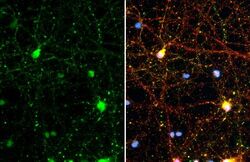
- Experimental details
- Immunofluorescent analysis of Synapsin II in DIV10 rat E18 primary cortical neuron cells. Cells were fixed in 4% paraformaldehyde at RT for 15 min. Green: Synapsin II stained by Synapsin II monoclonal antibody (Product # MA5-47281) diluted at 1:500. Red: Tau, a axon marker, stained by Tau antibody diluted at 1:500.

Supportive validation
- Submitted by
- Invitrogen Antibodies (provider)
- Main image

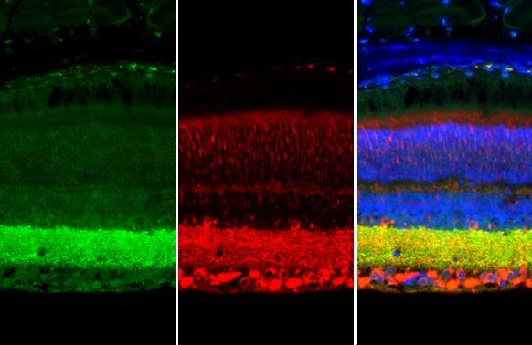
- Experimental details
- Immunohistochemistry analysis of Synapsin II in paraffin-embedded mouse eye. Green: Synapsin II stained by Synapsin II monoclonal antibody (Product # MA5-47281) diluted at 1:250. Red: beta Tubulin 3/ Tuj1, a cytoskeleton marker, stained by beta Tubulin 3/ Tuj1 antibody diluted at 1:500. Blue: Fluoroshield with DAPI. Antigen Retrieval: Citrate buffer, pH 6.0, 15 min.