Explore
 Validate
 Learn
LearnPA5-47746
antibody from Invitrogen Antibodies
Targeting: PLXNA2
FLJ11751, FLJ30634, KIAA0463, OCT, PLXN2
 Western blot
 Immunocytochemistry
Antibody data
- Antibody Data
- Antigen structure
- References [1]
- Comments [0]
- Validations
- Immunocytochemistry [5]
- Flow cytometry [2]
- Other assay [2]
Submit
Validation data
Reference
Comment
Report error
- Product number
- PA5-47746 - Provider product page

- Provider
- Invitrogen Antibodies
- Product name
- Plexin A2 Polyclonal Antibody
- Antibody type
- Polyclonal
- Antigen
- Recombinant full-length protein
- Description
- In direct ELISAs, less than 1% cross-reactivity with recombinant mouse (rm) Plexin A1, rmPlexin A3, and recombinant human Plexin B1 is observed. Reconstitute at 0.2 mg/mL in sterile PBS.
- Reactivity
- Human, Mouse, Rat
- Host
- Goat
- Isotype
- IgG
- Vial size
- 100 μg
- Concentration
- 0.2 mg/mL
- Storage
- -20°C, Avoid Freeze/Thaw Cycles
Submitted references PLXNA2 and LRRC40 as candidate genes in autism spectrum disorder.
Pijuan J, Ortigoza-Escobar JD, Ortiz J, Alcalá A, Calvo MJ, Cubells M, Hernando-Davalillo C, Palau F, Hoenicka J
Autism research : official journal of the International Society for Autism Research 2021 Jun;14(6):1088-1100
Autism research : official journal of the International Society for Autism Research 2021 Jun;14(6):1088-1100
No comments: Submit comment
Supportive validation
- Submitted by
- Invitrogen Antibodies (provider)
- Main image

- Experimental details
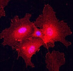
- Immunocytochemical analysis of Plexin A2 was detected in immersion fixed HUVEC human umbilical vein endothelial cells using Goat Anti-human/mouse/Rat Plexin A2 Antigen Affinity-purified Polyclonal Antibody (Product # PA5-47746) at 10 µg/mL for 3 hours at room temperature. Cells were stained using the 557-conjugated Anti-Goat IgG Secondary Antibody (re and counterstained with DAPI (blue). Specific staining was localized to cell surfaces and cytoplasm.

- Submitted by
- Invitrogen Antibodies (provider)
- Main image

- Experimental details
- Immunocytochemistry analysis of Plexin A2 in immersion fixed HUVEC human umbilical vein endothelial cells. Samples were incubated in Plexin A2 polyclonal antibody (Product # PA5-47746) using a dilution of 10 µg/mL for 3 hours at room temperature followed by NorthernLights™ 557-conjugated Anti-Goat IgG Secondary Antibody (red) and counterstained with DAPI (blue). Specific staining was localized to cell surfaces and cytoplasm.

- Submitted by
- Invitrogen Antibodies (provider)
- Main image

- Experimental details
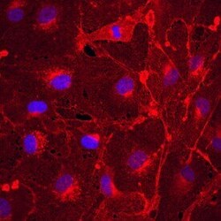
- Immunocytochemistry analysis of Plexin A2 in immersion fixed bEnd.3 mouse endothelioma cell line. Samples were incubated in Plexin A2 polyclonal antibody (Product # PA5-47746) using a dilution of 10 µg/mL for 3 hours at room temperature followed by NorthernLights™ 557-conjugated Anti-Goat IgG Secondary Antibody (red) and counterstained with DAPI (blue). Specific staining was localized to cell surfaces and cytoplasm.

- Submitted by
- Invitrogen Antibodies (provider)
- Main image
- Experimental details
- Immunocytochemistry analysis of Plexin A2 in immersion fixed HUVEC human umbilical vein endothelial cells. Samples were incubated in Plexin A2 polyclonal antibody (Product # PA5-47746) using a dilution of 10 µg/mL for 3 hours at room temperature followed by NorthernLights™ 557-conjugated Anti-Goat IgG Secondary Antibody (red) and counterstained with DAPI (blue). Specific staining was localized to cell surfaces and cytoplasm.

- Submitted by
- Invitrogen Antibodies (provider)
- Main image
- Experimental details
- Immunocytochemistry analysis of Plexin A2 in immersion fixed bEnd.3 mouse endothelioma cell line. Samples were incubated in Plexin A2 polyclonal antibody (Product # PA5-47746) using a dilution of 10 µg/mL for 3 hours at room temperature followed by NorthernLights™ 557-conjugated Anti-Goat IgG Secondary Antibody (red) and counterstained with DAPI (blue). Specific staining was localized to cell surfaces and cytoplasm.

Supportive validation
- Submitted by
- Invitrogen Antibodies (provider)
- Main image

- Experimental details
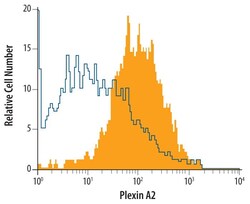
- Flow cytometry of Plexin A2 in bEnd.3 mouse endothelioma cell line. Samples were incubated in Plexin A2 polyclonal antibody (Product # PA5-47746) or isotype control antibody followed by Allophycocyanin-conjugated Anti-Goat IgG Secondary Antibody.

- Submitted by
- Invitrogen Antibodies (provider)
- Main image
- Experimental details
- Flow cytometry of Plexin A2 in bEnd.3 mouse endothelioma cell line. Samples were incubated in Plexin A2 polyclonal antibody (Product # PA5-47746) or isotype control antibody followed by Allophycocyanin-conjugated Anti-Goat IgG Secondary Antibody.

Supportive validation
- Submitted by
- Invitrogen Antibodies (provider)
- Main image
- Experimental details
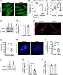
- 3 FIGURE PlxnA2 and LRRC40 variants affect the function of these proteins. (a) PlxnA2 subcellular localization in control and patient fibroblasts. (b) Percentage of nuclear/cytoplasmic or cytoplasmic localization patterns (box plot represents 25 th percentile, median, and 75 th percentile. The whiskers extend to the minimum and maximum values; n = 3 independent biological replicates; at least 100 random cells per experiment). Mantel-Haenszel chi 2 test (*** p

- Submitted by
- Invitrogen Antibodies (provider)
- Main image
- Experimental details
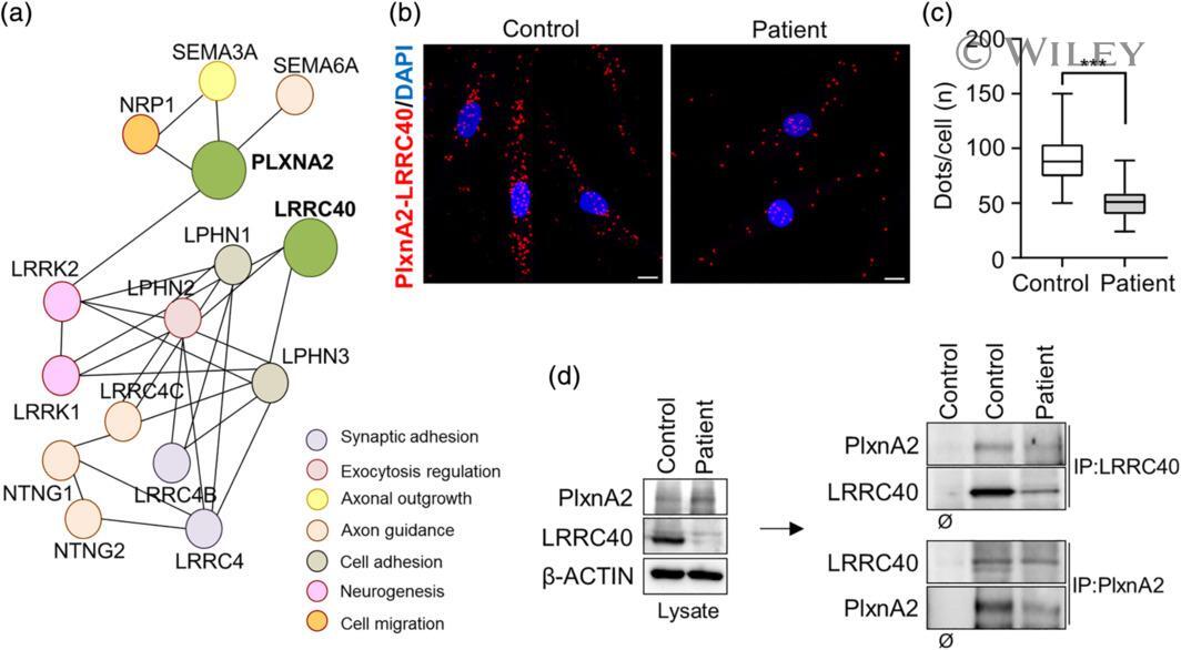
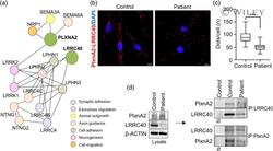
- 4 FIGURE PlxnA2 interacts with LRRC40. (a) Schematic representation of protein interaction network based on STRING software. (b) PLA of control and patient fibroblasts using alpha-PlxnA2 and alpha-LRRC40. (c) Quantification of PLA dots per cell (box plots represent 25 th percentile, median, 75 th percentile. The whiskers extend to the minimum and maximum values; n = 3 independent biological replicates; n = 60 cells). Mann-Whitney U -test (*** p