Explore
 Validate
 Learn
Learn Western blot
Antibody data
- Antibody Data
- Antigen structure
- References [2]
- Comments [0]
- Validations
- Western blot [2]
- Immunocytochemistry [2]
- Flow cytometry [1]
Submit
Validation data
Reference
Comment
Report error
- Product number
- AF5486 - Provider product page

- Provider
- R&D Systems
- Product name
- Human/Mouse/Rat Plexin A2 Antibody
- Antibody type
- Polyclonal
- Description
- Immunogen affinity purified. Detects mouse and rat Plexin A2 in direct ELISAs and Western blots. In direct ELISAs, less than 1% cross-reactivity with recombinant mouse (rm) Plexin A1, rmPlexin A3, and recombinant human Plexin B1 is observed.
- Reactivity
- Human, Mouse, Rat
- Host
- Goat
- Conjugate
- Unconjugated
- Antigen sequence
P70207- Isotype
- IgG
- Vial size
- 100 ug
- Concentration
- LYOPH
- Storage
- Use a manual defrost freezer and avoid repeated freeze-thaw cycles. 12 months from date of receipt, -20 to -70 °C as supplied. 1 month, 2 to 8 °C under sterile conditions after reconstitution. 6 months, -20 to -70 °C under sterile conditions after reconstitution.
Submitted references Sympathetic nerve repulsion inhibited by designer molecules in vitro and role in experimental arthritis.
Neural crest-derived SEMA3C activates endothelial NRP1 for cardiac outflow tract septation.
Kunath J, Delaroque N, Szardenings M, Neundorf I, Straub RH
Life sciences 2017 Jan 1;168:47-53
Life sciences 2017 Jan 1;168:47-53
Neural crest-derived SEMA3C activates endothelial NRP1 for cardiac outflow tract septation.
Plein A, Calmont A, Fantin A, Denti L, Anderson NA, Scambler PJ, Ruhrberg C
The Journal of clinical investigation 2015 Jul 1;125(7):2661-76
The Journal of clinical investigation 2015 Jul 1;125(7):2661-76
No comments: Submit comment
Supportive validation
- Submitted by
- R&D Systems (provider)
- Main image
- Experimental details
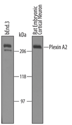
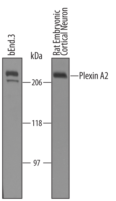
- Detection of Mouse and Rat Plexin A2 by Western Blot. Western blot shows lysates of bEnd.3 mouse endothelioma cell line and rat embryonic cortical neuron cells. PVDF Membrane was probed with 1 µg/mL of Goat Anti-Human/Mouse/Rat Plexin A2 Antigen Affinity-purified Polyclonal Antibody (Catalog # AF5486) followed by HRP-conjugated Anti-Goat IgG Secondary Antibody (Catalog # HAF019). A specific band was detected for Plexin A2 at approximately 210 kDa (as indicated). This experiment was conducted under reducing conditions and using Immunoblot Buffer Group 8.
- Submitted by
- R&D Systems (provider)
- Main image

- Experimental details
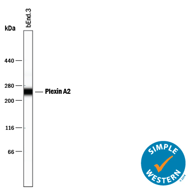
- Detection of Mouse Plexin A2 by Simple WesternTM. Simple Western lane view shows lysates of bEnd.3 mouse endothelioma cell line, loaded at 0.2 mg/mL. A specific band was detected for Plexin A2 at approximately 246 kDa (as indicated) using 10 µg/mL of Goat Anti-Human/Mouse/Rat Plexin A2 Antigen Affinity-purified Polyclonal Antibody (Catalog # AF5486) followed by 1:50 dilution of HRP-conjugated Anti-Goat IgG Secondary Antibody (Catalog # HAF109). This experiment was conducted under reducing conditions and using the 66-440 kDa separation system.
Supportive validation
- Submitted by
- R&D Systems (provider)
- Main image
- Experimental details
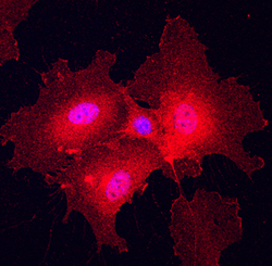
- Plexin A2 in HUVEC Human Cells. Plexin A2 was detected in immersion fixed HUVEC human umbilical vein endothelial cells using Goat Anti-Human/Mouse/Rat Plexin A2 Antigen Affinity-purified Polyclonal Antibody (Catalog # AF5486) at 10 µg/mL for 3 hours at room temperature. Cells were stained using the NorthernLights™ 557-conjugated Anti-Goat IgG Secondary Antibody (red; Catalog # NL001) and counterstained with DAPI (blue). Specific staining was localized to cell surfaces and cytoplasm. View our protocol for Fluorescent ICC Staining of Cells on Coverslips.

- Submitted by
- R&D Systems (provider)
- Main image
- Experimental details
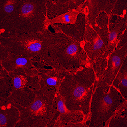
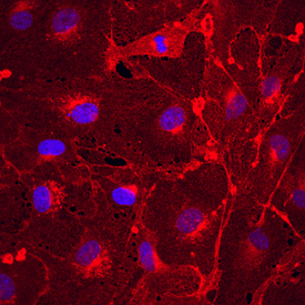
- Plexin A2 in bEnd.3 Mouse Cell Line. Plexin A2 was detected in immersion fixed bEnd.3 mouse endothelioma cell line using Goat Anti-Human/Mouse/Rat Plexin A2 Antigen Affinity-purified Polyclonal Antibody (Catalog # AF5486) at 10 µg/mL for 3 hours at room temperature. Cells were stained using the NorthernLights™ 557-conjugated Anti-Goat IgG Secondary Antibody (red; Catalog # NL001) and counterstained with DAPI (blue). Specific staining was localized to cell surfaces and cytoplasm. View our protocol for Fluorescent ICC Staining of Cells on Coverslips.
Supportive validation
- Submitted by
- R&D Systems (provider)
- Main image

- Experimental details
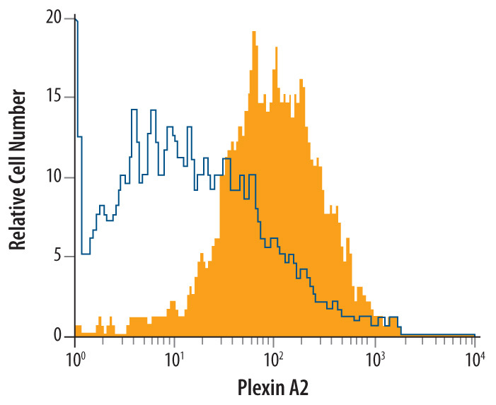
- Detection of Plexin A2 in bEnd.3 Mouse Cell Line by Flow Cytometry. bEnd.3 mouse endothelioma cell line was stained with Goat Anti-Human/Mouse/Rat Plexin A2 Antigen Affinity-purified Polyclonal Antibody (Catalog # AF5486, filled histogram) or isotype control antibody (Catalog # AB-108-C, open histogram), followed by Allophycocyanin-conjugated Anti-Goat IgG Secondary Antibody (Catalog # F0108).