Explore
 Validate
 Learn
LearnPB9329
antibody from Boster Biological Technology
Targeting: CACNA1D
CACH3, CACN4, CACNL1A2, Cav1.3, CCHL1A2
 Western blot
 Immunocytochemistry
Antibody data
- Antibody Data
- Antigen structure
- References [0]
- Comments [0]
- Validations
- Western blot [1]
Submit
Validation data
Reference
Comment
Report error
- Product number
- PB9329 - Provider product page

- Provider
- Boster Biological Technology
- Product name
- Anti-CaV1.3/CACNA1D Antibody Picoband™
- Antibody type
- Polyclonal
- Description
- Polyclonal antibody for CAV1.3/CACNA1D detection. Host: Rabbit.Size: 100μg/vial. Tested applications: WB. Reactive species: Human. CAV1.3/CACNA1D information: Molecular Weight: 245141 MW; Subcellular Localization: Membrane ; Multi-pass membrane protein ; Tissue Specificity: Expressed in pancreatic islets and in brain, where it has been seen in cerebral cortex, hippocampus, basal ganglia, habenula and thalamus. Expressed in the small cell lung carcinoma cell line SCC-9. No expression in skeletal muscle.
- Reactivity
- Human, Mouse, Rat
- Host
- Rabbit
- Vial size
- 100μg/vial
- Concentration
- Add 0.2ml of distilled water will yield a concentration of 500ug/ml.
- Storage
- At -20°C for one year. After reconstitution, at 4°C for one month. It can also be aliquoted and stored frozen at -20°C for a longer time. Avoid repeated freezing and thawing.
- Handling
- Add 0.2ml of distilled water will yield a concentration of 500ug/ml.
No comments: Submit comment
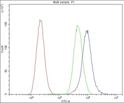
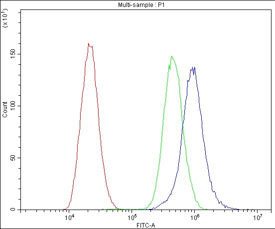
Supportive validation
- Submitted by
- Boster Biological Technology (provider)
- Main image
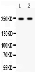
- Experimental details
- Western blot analysis of CAV1.3 using anti-CAV1.3 antibody (PB9329). Electrophoresis was performed on a 5-20% SDS-PAGE gel at 70V (Stacking gel) / 90V (Resolving gel) for 2-3 hours. The sample well of each lane was loaded with 50ug of sample under reducing conditions. Lane 1: Rat Brain Tissue Lysate, Lane 2: Mouse Brain Tissue Lysate. After Electrophoresis, proteins were transferred to a Nitrocellulose membrane at 150mA for 50-90 minutes. Blocked the membrane with 5% Non-fat Milk/ TBS for 1.5 hour at RT. The membrane was incubated with rabbit anti-CAV1.3 antigen affinity purified polyclonal antibody (Catalog # PB9329) at 0.5 μg/mL overnight at 4°C, then washed with TBS-0.1%Tween 3 times with 5 minutes each and probed with a goat anti-rabbit IgG-HRP secondary antibody at a dilution of 1:10000 for 1.5 hour at RT. The signal is developed using an Enhanced Chemiluminescent detection (ECL) kit (Catalog # EK1002) with Tanon 5200 system. A specific band was detected for CAV1.3 at approximately 245KD. The expected band size for CAV1.3 is at 245KD.
- Additional image