Explore
 Validate
 Learn
LearnACC-311-200UL
antibody from Invitrogen Antibodies
Targeting: CACNA1D
CACH3, CACN4, CACNL1A2, Cav1.3, CCHL1A2
 Western blot
Antibody data
- Antibody Data
- Antigen structure
- References [0]
- Comments [0]
- Validations
- Western blot [1]
- Immunocytochemistry [2]
- Immunohistochemistry [1]
- Flow cytometry [1]
Submit
Validation data
Reference
Comment
Report error
- Product number
- ACC-311-200UL - Provider product page

- Provider
- Invitrogen Antibodies
- Product name
- CaV1.3 (CACNA1D) (extracellular) Polyclonal Antibody
- Antibody type
- Polyclonal
- Antigen
- Other
- Reactivity
- Human, Mouse, Rat
- Host
- Rabbit
- Isotype
- IgG
- Vial size
- 200 µL
- Concentration
- 0.9 mg/mL
- Storage
- -20° C, Avoid Freeze/Thaw Cycles
No comments: Submit comment
Supportive validation
- Submitted by
- Invitrogen Antibodies (provider)
- Main image
- Experimental details
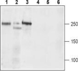
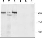
- Western blot analysis of rat brain (lanes 1 and 4), mouse brain (lanes 2 and 5), and ratC6 brain Glioma (lanes 3 and 6) lysates: - 1-3. Anti-CaV1.3 (CACNA1D) (extracellular) Antibody (#ACC-311), (1:200).4-6. Anti-CaV1.3 (CACNA1D) (extracellular) Antibody , preincubated with Cav1.3/CACNA1D (extracellular) Blocking Peptide (#BLP-CC311).
Supportive validation
- Submitted by
- Invitrogen Antibodies (provider)
- Main image

- Experimental details
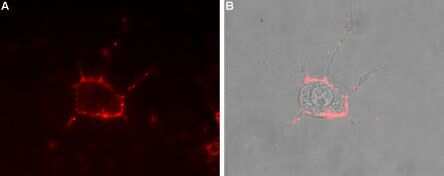
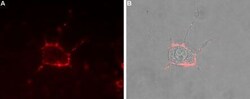
- Expression of CaV1.3 in rat PC12 cells - Cell surface detection of CaV1.3 in intact living rat pheochromocytoma (PC12) cells. A. Extracellular staining of cells using Anti-CaV1.3 (CACNA1D) (extracellular) Antibody (#ACC-311), (1:50), (red). B. Merge of A with the live view of the cell.
- Submitted by
- Invitrogen Antibodies (provider)
- Main image
- Experimental details
- Expression of CaV1.3 in rat PC12 cells - Cell surface detection of CaV1.3 in intact living rat pheochromocytoma (PC12) cells. A. Extracellular staining of cells using Anti-CaV1.3 (CACNA1D) (extracellular) Antibody (#ACC-311), (1:50), (red). B. Merge of A with the live view of the cell.

Supportive validation
- Submitted by
- Invitrogen Antibodies (provider)
- Main image
- Experimental details
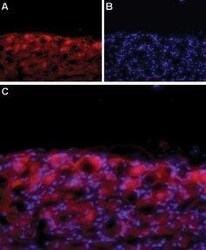
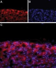
- Expression of CaV1.3 in rat DRG - Immunohistochemical staining of adult rat dorsal root ganglion (DRG) using Anti-CaV1.3 (CACNA1D) (extracellular) Antibody (#ACC-311). A. CaV1.3 labeling (red) appears in the cell bodies of the DRG. B. Nuclear staining using DAPI as the counterstain. C. Merged image of A and B.
Supportive validation
- Submitted by
- Invitrogen Antibodies (provider)
- Main image
- Experimental details
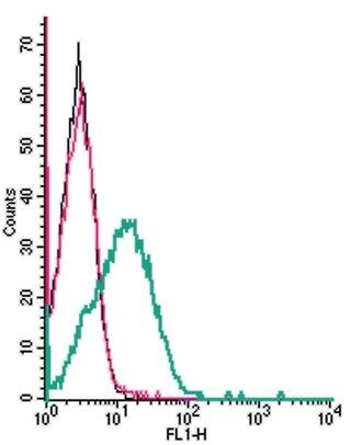
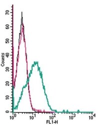
- Cell surface detection ofCav1.3by indirect flow cytometry in live intacthumanJurkatT-cellleukemiacells: - (black line) cells. (red) Cells+ goat- Anti-rabbit-FITC. (green) Cells + Anti-CaV1.3 (CACNA1D) (extracellular) Antibody (#ACC-311), (5μg) + goat- Anti-rabbit-FITC.