Explore
 Validate
 Learn
Learn Western blot
 Immunocytochemistry
Antibody data
- Antibody Data
- Antigen structure
- References [1]
- Comments [0]
- Validations
- Immunocytochemistry [2]
- Immunohistochemistry [2]
- Other assay [1]
Submit
Validation data
Reference
Comment
Report error
- Product number
- PA5-78052 - Provider product page

- Provider
- Invitrogen Antibodies
- Product name
- MAP1B Polyclonal Antibody
- Antibody type
- Polyclonal
- Antigen
- Synthetic peptide
- Description
- Positive Control: SK-N-AS, C8D30 Predicted Reactivity: Mouse (100%) Store product as a concentrated solution. Centrifuge briefly prior to opening the vial.
- Reactivity
- Human, Mouse, Rat
- Host
- Rabbit
- Isotype
- IgG
- Vial size
- 100 μL
- Concentration
- 1.28 mg/mL
- Storage
- Store at 4°C short term. For long term storage, store at -20°C, avoiding freeze/thaw cycles.
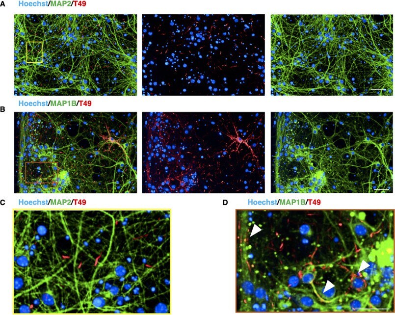
Submitted references Quantitative propagation of assembled human Tau from Alzheimer's disease brain in microfluidic neuronal cultures.
Katsikoudi A, Ficulle E, Cavallini A, Sharman G, Guyot A, Zagnoni M, Eastwood BJ, Hutton M, Bose S
The Journal of biological chemistry 2020 Sep 11;295(37):13079-13093
The Journal of biological chemistry 2020 Sep 11;295(37):13079-13093
No comments: Submit comment
Supportive validation
- Submitted by
- Invitrogen Antibodies (provider)
- Main image

- Experimental details
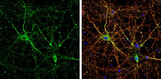
- MAP1B Polyclonal Antibody detects MAP1B protein by immunofluorescent analysis. Sample: DIV14 rat E18 primary cortical neurons were fixed in 4% paraformaldehyde at RT for 15 min. Green: MAP1B protein stained by MAP1B Polyclonal Antibody (Product # PA5-78052) diluted at 1:500. Red: beta Tubulin 3/ Tuj1, stained by beta Tubulin 3/ Tuj1 antibody [GT1338] diluted at 1:500. Blue: Fluoroshield with DAPI .
- Submitted by
- Invitrogen Antibodies (provider)
- Main image

- Experimental details
- MAP1B Polyclonal Antibody detects MAP1B protein by immunofluorescent analysis. Sample: DIV14 rat E18 primary cortical neurons were fixed in 4% paraformaldehyde at RT for 15 min. Green: MAP1B protein stained by MAP1B Polyclonal Antibody (Product # PA5-78052) diluted at 1:500. Red: beta Tubulin 3/ Tuj1, stained by beta Tubulin 3/ Tuj1 antibody [GT1338] diluted at 1:500. Blue: Fluoroshield with DAPI .

Supportive validation
- Submitted by
- Invitrogen Antibodies (provider)
- Main image
- Experimental details
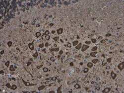
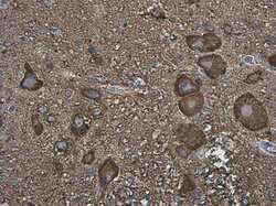
- MAP1B Polyclonal Antibody detects MAP1B protein at cytoplasm in mouse brain by immunohistochemical analysis. Sample: Paraffin-embedded mouse brain. MAP1B Polyclonal Antibody (Product # PA5-78052) diluted at 1:500. Antigen Retrieval: Citrate buffer, pH 6.0, 15 min.
- Submitted by
- Invitrogen Antibodies (provider)
- Main image
- Experimental details
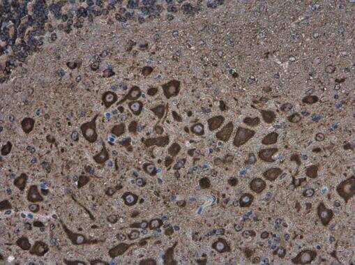
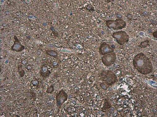
- MAP1B Polyclonal Antibody detects MAP1B protein at cytoplasm in rat brain by immunohistochemical analysis. Sample: Paraffin-embedded rat brain. MAP1B Polyclonal Antibody (Product # PA5-78052) diluted at 1:500. Antigen Retrieval: Citrate buffer, pH 6.0, 15 min.
Supportive validation
- Submitted by
- Invitrogen Antibodies (provider)
- Main image
- Experimental details
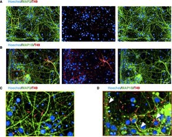
- Figure 2. T49-positive inclusions are localized in the axons and soma of RCNs. A-D , sparse co-localization between the T49 neuritic thread-like inclusions in RCNs and MAP2, a specific dendritic marker ( A and zoomed image in C ), but good co-localization with MAP1B, a neuronal cytoskeleton marker ( B and zoomed image in D ), indicating that the inclusions observed after seeding with hAD seed are not dendritic but somatic and axonal. Arrowheads in the D indicate co-localization between MAP1B and T49. Images were acquired with Opera Phenix and 20x objective. Figure bar , 50 mum; zoomed image bar , 25 mum.