 Explore
 Validate
 Learn
LearnHPA024210
antibody from Atlas Antibodies
Targeting: NCOA3
ACTR, AIB1, bHLHe42, CAGH16, KAT13B, p/CIP, RAC3, SRC-3, SRC3, TNRC16, TRAM-1
 Western blot
 Immunocytochemistry
Antibody data
- Antibody Data
- Antigen structure
- References [2]
- Comments [0]
- Validations
- Immunocytochemistry [1]
- Chromatin Immunoprecipitation [1]
Submit
Validation data
Reference
Comment
Report error
- Product number
- HPA024210 - Provider product page

- Provider
- Atlas Antibodies
- Proper citation
- Atlas Antibodies Cat#HPA024210, RRID:AB_10602011
- Product name
- Anti-NCOA3
- Antibody type
- Polyclonal
- Description
- Polyclonal Antibody against Human NCOA3, Gene description: nuclear receptor coactivator 3, Alternative Gene Names: ACTR, AIB1, bHLHe42, CAGH16, KAT13B, p/CIP, RAC3, SRC-3, SRC3, TNRC16, TRAM-1, Validated applications: WB, IHC, ICC, ChIP, Uniprot ID: Q9Y6Q9, Storage: Store at +4°C for short term storage. Long time storage is recommended at -20°C.
- Reactivity
- Human
- Host
- Rabbit
- Conjugate
- Unconjugated
- Isotype
- IgG
- Vial size
- 100 µl
- Concentration
- 0.3 mg/ml
- Storage
- Store at +4°C for short term storage. Long time storage is recommended at -20°C.
- Handling
- The antibody solution should be gently mixed before use.
Submitted references The High Expression of the microRNA 17–92 Cluster and its Paralogs, and the Downregulation of the Target Gene PTEN, Is Associated with Primary Cutaneous B-Cell Lymphoma Progression
The High Expression of the microRNA 17–92 Cluster and its Paralogs, and the Downregulation of the Target Gene PTEN, Is Associated with Primary Cutaneous B-Cell Lymphoma Progression
Battistella M, Romero M, Castro-Vega L, Gapihan G, Bouhidel F, Bagot M, Feugeas J, Janin A
Journal of Investigative Dermatology 2015;135(6):1659-1667
Journal of Investigative Dermatology 2015;135(6):1659-1667
The High Expression of the microRNA 17–92 Cluster and its Paralogs, and the Downregulation of the Target Gene PTEN, Is Associated with Primary Cutaneous B-Cell Lymphoma Progression
Battistella M, Romero M, Castro-Vega L, Gapihan G, Bouhidel F, Bagot M, Feugeas J, Janin A
Journal of Investigative Dermatology 2015 June;135(6):1659-1667
Journal of Investigative Dermatology 2015 June;135(6):1659-1667
No comments: Submit comment
Supportive validation
- Submitted by
- Atlas Antibodies (provider)
- Main image
- Experimental details
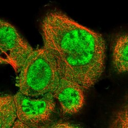
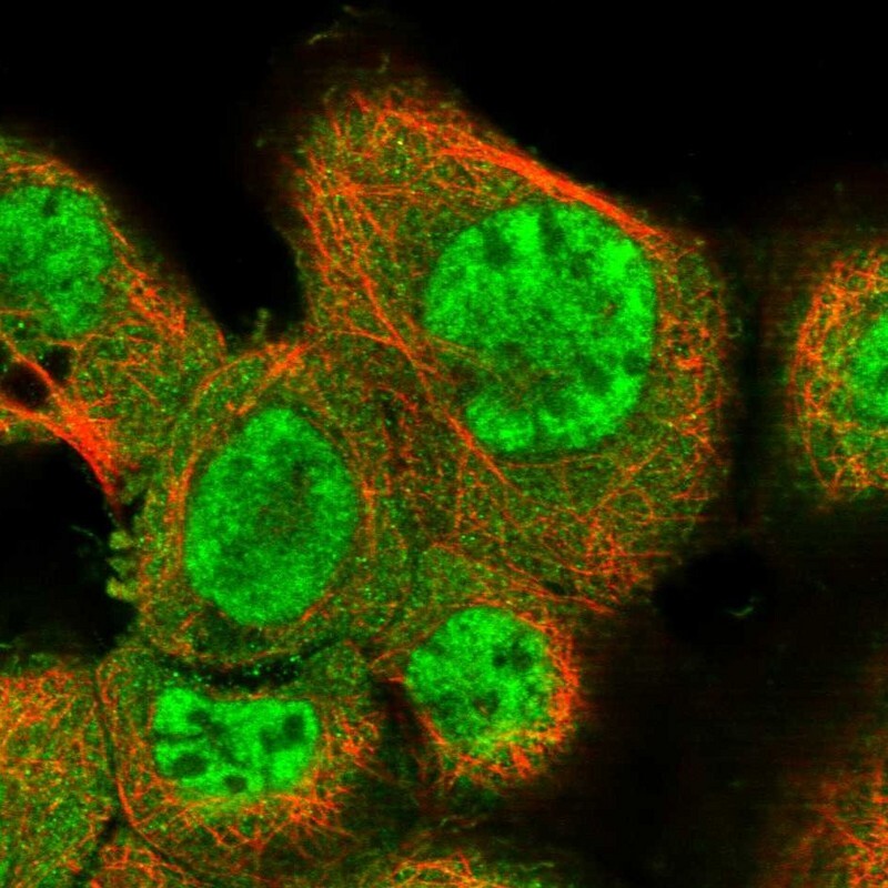
- Immunofluorescent staining of human cell line A-431 shows localization to nucleoplasm & cytosol.
- Sample type
- Human
Supportive validation
- Submitted by
- Atlas Antibodies (provider)
- Main image
- Experimental details
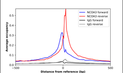
- ChIP-Exo-Seq composite graph for Anti-NCOA3 (HPA024210, Lot 000000578) tested in K562 cells. Strand-specific reads (blue: forward, red: reverse) and IgG controls (black: forward, grey: reverse) are plotted against the distance from a composite set of reference binding sites. The antibody exhibits robust target enrichment compared to a non-specific IgG control and precisely reveals its structural organization around the binding site. Data generated by Prof. B. F. Pugh´s Lab at Cornell University.
