Explore
 Validate
 Learn
Learn Western blot
Antibody data
- Antibody Data
- Antigen structure
- References [0]
- Comments [0]
- Validations
- Western blot [4]
- Immunohistochemistry [13]
Submit
Validation data
Reference
Comment
Report error
- Product number
- MA5-25223 - Provider product page

- Provider
- Invitrogen Antibodies
- Product name
- EPHX2 Monoclonal Antibody (OTI1H5)
- Antibody type
- Monoclonal
- Antigen
- Recombinant full-length protein
- Reactivity
- Human
- Host
- Mouse
- Isotype
- IgG
- Antibody clone number
- OTI1H5
- Vial size
- 100 µL
- Concentration
- 1 mg/mL
- Storage
- -20° C, Avoid Freeze/Thaw Cycles
No comments: Submit comment
Supportive validation
- Submitted by
- Invitrogen Antibodies (provider)
- Main image
- Experimental details
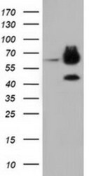
- Western blot analysis of EPHX2 in HEK293T cells in untransfected (Left lane) and transfected (Right lane) samples using 5 µg per lane. The samples were separated by SDS-PAGE and probed with EPHX2 (Product # MA5-25223) monoclonal antibody.
- Submitted by
- Invitrogen Antibodies (provider)
- Main image
- Experimental details
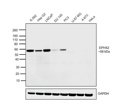
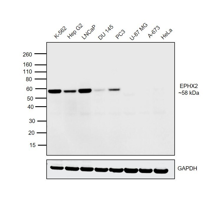
- Western blot was performed using Anti-EPHX2 Monoclonal Antibody (OTI1H5) (Product # MA5-25223) and a 58 kDa band corresponding to bifunctional Epoxide Hydrolase 2 (EPHX2) was observed across cell lines tested except U-87 MG, A-673 and HeLa. Whole cell extracts (30 µg lysate) of K-562 (Lane 1), Hep G2 (Lane 2), LNCaP (Lane 3), DU 145 (Lane 4), PC-3 (Lane 5), U-87 MG (Lane 6), A-673 (Lane 7) and HeLa (Lane 8) were electrophoresed using NuPAGE™ 4-12% Bis-Tris Protein Gel (Product # NP0321BOX), 10 well. Resolved proteins were then transferred onto a nitrocellulose membrane (Product # IB23001) by iBlot® 2 Dry Blotting System (Product # IB21001). The blot was probed with the primary antibody (1:1000 dilution) and detected by chemiluminescence with Goat anti-Mouse IgG (H+L) Superclonal™ Recombinant Secondary Antibody, HRP (Product # A28177, 1:20,000 dilution) using the iBright™ FL1500 Imaging System (Product # A44115). Chemiluminescent detection was performed using SuperSignal™ West Pico PLUS Chemiluminescent Substrate (Product # 34580).
- Submitted by
- Invitrogen Antibodies (provider)
- Main image

- Experimental details
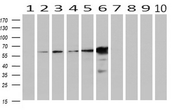
- Western blot analysis of EPHX2 in human tissue (1: Testis; 2: Omentum; 3: Uterus; 4: Breast; 5: Brain; 6: Liver; 7: Ovary; 8: Thyroid gland; 9: colon;10: spleen) samples using 10 µg per lane. Samples were separated by SDS-PAGE and probed with EPHX2 (Product # MA5-25223) monoclonal antibody at a dilution of 1:200.
- Submitted by
- Invitrogen Antibodies (provider)
- Main image
- Experimental details
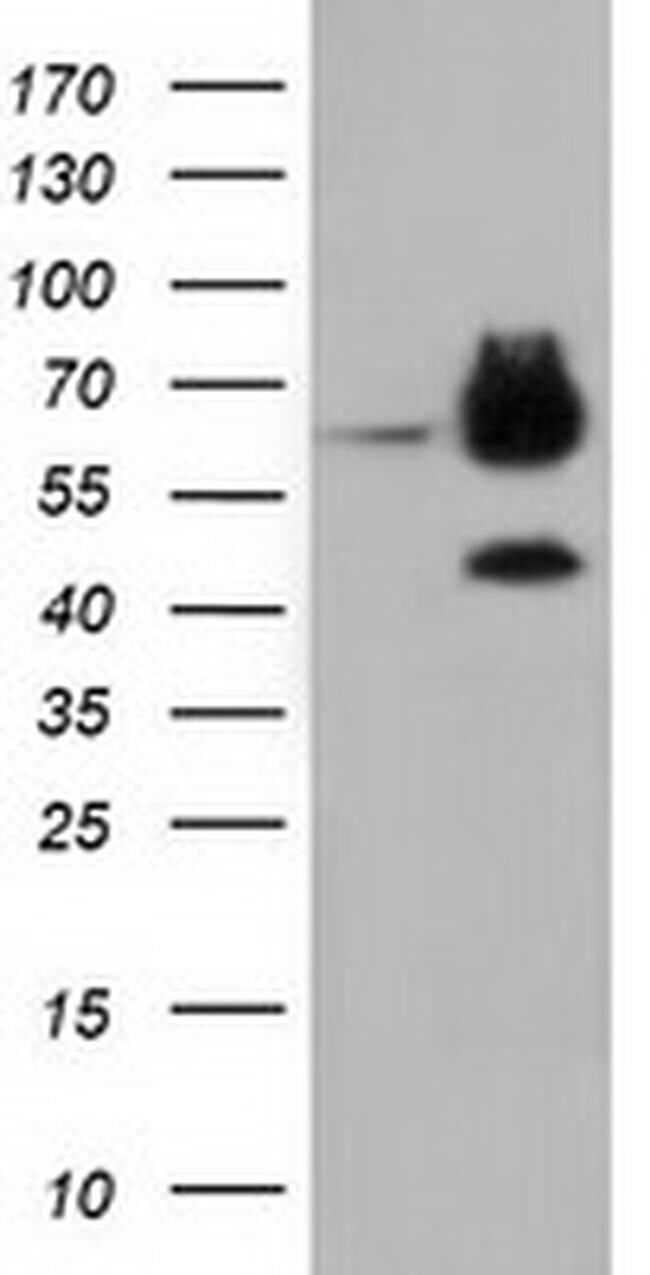
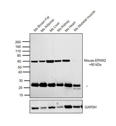
- Western blot was performed using Anti-EPHX2 Monoclonal Antibody (OTI1H5) (Product # MA5-25223) and a 60 kDa band corresponding to bifunctional Epoxide Hydrolase 2 (EPHX2) was observed across mouse tissues tested except skeletal muscle. Whole cell extracts (30 µg lysate) of Mouse Brown Fat (Lane 1), Mouse Adipose (Lane 2), Mouse Liver (Lane 3), Mouse Kidney (Lane 4), Mouse Heart (Lane 5) and Mouse Skeletal Muscle (Lane 6) were electrophoresed using NuPAGE™ 4-12% Bis-Tris Protein Gel (Product # NP0321BOX), 10 well. Resolved proteins were then transferred onto a nitrocellulose membrane (Product # IB23001) by iBlot® 2 Dry Blotting System (Product # IB21001). The blot was probed with the primary antibody (1:1000 dilution) and detected by chemiluminescence with Goat anti-Mouse IgG (H+L) Superclonal™ Recombinant Secondary Antibody, HRP (Product # A28177, 1:20,000 dilution) using the iBright™ FL1500 Imaging System (Product # A44115). Chemiluminescent detection was performed using SuperSignal™ West Pico PLUS Chemiluminescent Substrate (Product # 34580). A band corresponding to tissue IgG was observed around ~25 kDa.

Supportive validation
- Submitted by
- Invitrogen Antibodies (provider)
- Main image
- Experimental details
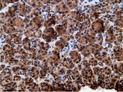
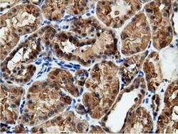
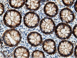
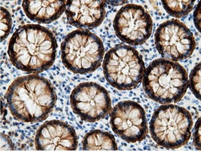
- Immunohistochemistry was performed on paraffin-embedded human pancreas tissue. To expose target proteins, 10mM citric buffer, pH6.0, 100°C for 10min was used. Following antigen retrieval, tissues were probed with a EPHX2 monoclonal antibody (Product # MA5-25223).
- Submitted by
- Invitrogen Antibodies (provider)
- Main image
- Experimental details
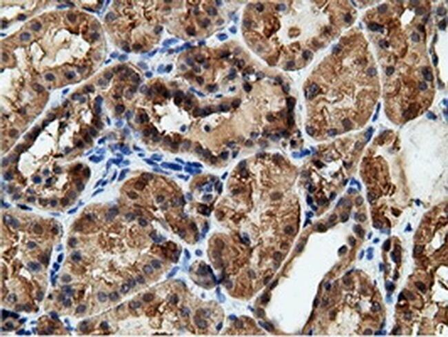
- Immunohistochemistry was performed on paraffin-embedded human kidney tissue. To expose target proteins, 10mM citric buffer, pH6.0, 100°C for 10min was used. Following antigen retrieval, tissues were probed with a EPHX2 monoclonal antibody (Product # MA5-25223).
- Submitted by
- Invitrogen Antibodies (provider)
- Main image

- Experimental details
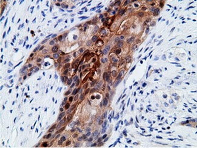
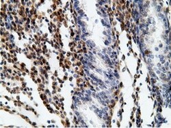
- Immunohistochemistry was performed on paraffin-embedded adenocarcinoma of human colon tissue. To expose target proteins, 10mM citric buffer, pH6.0, 100°C for 10min was used. Following antigen retrieval, tissues were probed with a EPHX2 monoclonal antibody (Product # MA5-25223).

- Submitted by
- Invitrogen Antibodies (provider)
- Main image

- Experimental details
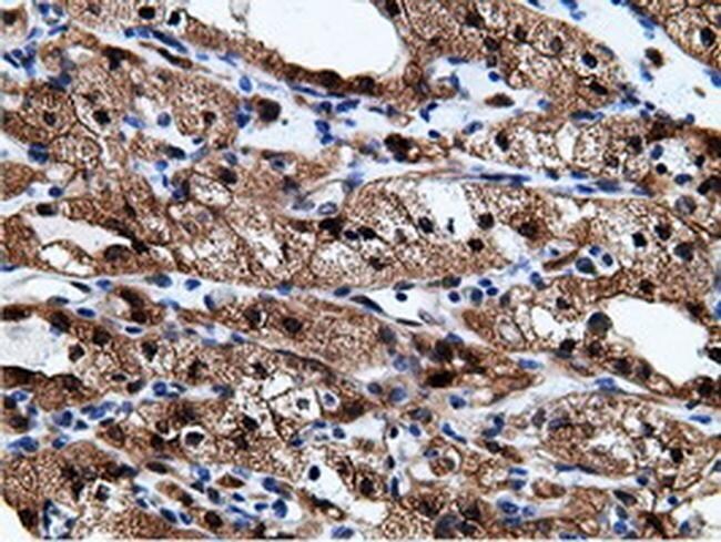
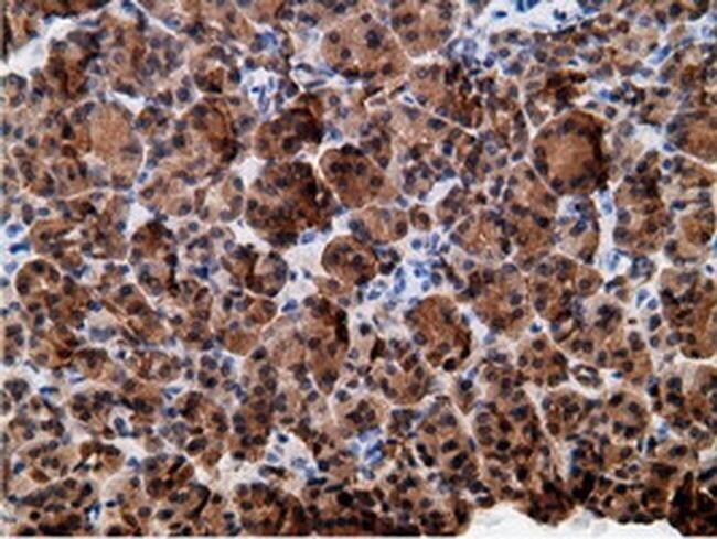
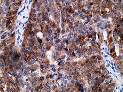
- Immunohistochemistry was performed on paraffin-embedded carcinoma of human pancreas tissue. To expose target proteins, 10mM citric buffer, pH6.0, 100°C for 10min was used. Following antigen retrieval, tissues were probed with a EPHX2 monoclonal antibody (Product # MA5-25223).
- Submitted by
- Invitrogen Antibodies (provider)
- Main image
- Experimental details
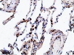
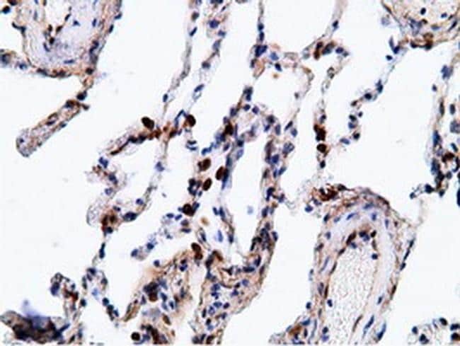
- Immunohistochemistry was performed on paraffin-embedded human lung tissue. To expose target proteins, 10mM citric buffer, pH6.0, 100°C for 10min was used. Following antigen retrieval, tissues were probed with a EPHX2 monoclonal antibody (Product # MA5-25223).
- Submitted by
- Invitrogen Antibodies (provider)
- Main image
- Experimental details
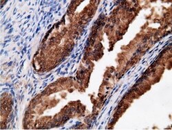
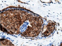
- Immunohistochemistry was performed on paraffin-embedded human prostate tissue. To expose target proteins, 10mM citric buffer, pH6.0, 100°C for 10min was used. Following antigen retrieval, tissues were probed with a EPHX2 monoclonal antibody (Product # MA5-25223).

- Submitted by
- Invitrogen Antibodies (provider)
- Main image

- Experimental details
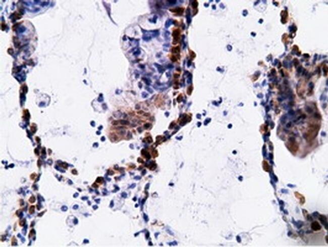
- Immunohistochemistry was performed on paraffin-embedded carcinoma of human lung tissue. To expose target proteins, 10mM citric buffer, pH6.0, 100°C for 10min was used. Following antigen retrieval, tissues were probed with a EPHX2 monoclonal antibody (Product # MA5-25223).
- Submitted by
- Invitrogen Antibodies (provider)
- Main image
- Experimental details
- Immunohistochemistry was performed on paraffin-embedded carcinoma of human bladder tissue. To expose target proteins, 10mM citric buffer, pH6.0, 100°C for 10min was used. Following antigen retrieval, tissues were probed with a EPHX2 monoclonal antibody (Product # MA5-25223).
- Submitted by
- Invitrogen Antibodies (provider)
- Main image
- Experimental details
- Immunohistochemistry was performed on paraffin-embedded adenocarcinoma of human breast tissue. To expose target proteins, 10mM citric buffer, pH6.0, 100°C for 10min was used. Following antigen retrieval, tissues were probed with a EPHX2 monoclonal antibody (Product # MA5-25223).

- Submitted by
- Invitrogen Antibodies (provider)
- Main image
- Experimental details
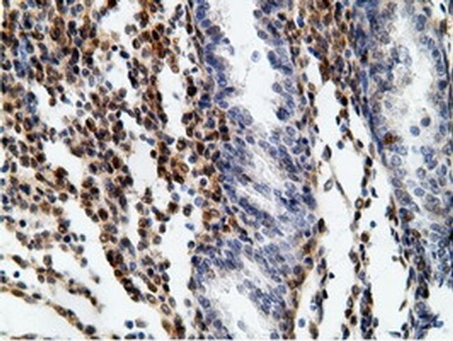
- Immunohistochemistry was performed on paraffin-embedded human colon tissue. To expose target proteins, 10mM citric buffer, pH6.0, 100°C for 10min was used. Following antigen retrieval, tissues were probed with a EPHX2 monoclonal antibody (Product # MA5-25223).
- Submitted by
- Invitrogen Antibodies (provider)
- Main image
- Experimental details
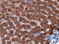
- Immunohistochemistry was performed on paraffin-embedded human liver tissue. To expose target proteins, 10mM citric buffer, pH6.0, 100°C for 10min was used. Following antigen retrieval, tissues were probed with a EPHX2 monoclonal antibody (Product # MA5-25223).

- Submitted by
- Invitrogen Antibodies (provider)
- Main image
- Experimental details
- Immunohistochemistry was performed on paraffin-embedded adenocarcinoma of human ovary tissue. To expose target proteins, 10mM citric buffer, pH6.0, 100°C for 10min was used. Following antigen retrieval, tissues were probed with a EPHX2 monoclonal antibody (Product # MA5-25223).

- Submitted by
- Invitrogen Antibodies (provider)
- Main image
- Experimental details
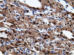
- Immunohistochemistry was performed on paraffin-embedded carcinoma of human kidney tissue. To expose target proteins, 10mM citric buffer, pH6.0, 100°C for 10min was used. Following antigen retrieval, tissues were probed with a EPHX2 monoclonal antibody (Product # MA5-25223).