Explore
 Validate
 Learn
Learn Western blot
 ELISA
ELISAAntibody data
- Antibody Data
- Antigen structure
- References [1]
- Comments [0]
- Validations
- Western blot [1]
- Immunohistochemistry [2]
Submit
Validation data
Reference
Comment
Report error
- Product number
- NBP2-21667 - Provider product page

- Provider
- Novus Biologicals
- Product name
- Rabbit Polyclonal Kinesin 5B Antibody
- Antibody type
- Polyclonal
- Description
- Delipidation and Defibrination.
- Reactivity
- Human
- Host
- Rabbit
- Vial size
- 0.1 ml
- Storage
- Store at -20C. Avoid freeze-thaw cycles.
Submitted references Glucocorticoid-mediated ER-mitochondria contacts reduce AMPA receptor and mitochondria trafficking into cell terminus via microtubule destabilization.
Choi GE, Oh JY, Lee HJ, Chae CW, Kim JS, Jung YH, Han HJ
Cell death & disease 2018 Nov 14;9(11):1137
Cell death & disease 2018 Nov 14;9(11):1137
No comments: Submit comment
Supportive validation
- Submitted by
- Novus Biologicals (provider)
- Main image

- Experimental details
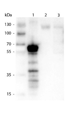
- Western Blot: Kinesin Antibody [NBP2-21667] - Rabbit anti-Kinesin 1 Antibody. Lane 1: 500 ng of truncated kinesin 1 protein. Lane 2: 20 ug of Mouse Brain lysate. Lane 3: 10 ug of Mouse Brain lysate. kinesin1 antibody at 1:1000 for overnight at 4C. Secondary antibody: Dye800 rabbit secondary antibody at 1:40,000 for 1hr at RT. Block: 5% Blocking Buffer1 hr at RT. Predicted/Observed size: 72 kDa, ~70 kDa for kinesin-1. Other band: degradation.
Supportive validation
- Submitted by
- Novus Biologicals (provider)
- Main image
- Experimental details
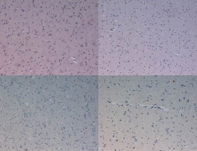
- Immunohistochemistry: Kinesin Antibody [NBP2-21667] - 40X in human brain at pH 6. Tissue: Human Brain.
- Submitted by
- Novus Biologicals (provider)
- Main image

- Experimental details
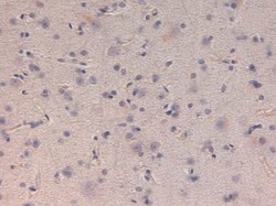
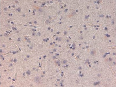
- Immunohistochemistry: Kinesin Antibody [NBP2-21667] - 40X in human brain at pH 6. Tissue: Human Brain.