 Explore
 Validate
 Learn
Learn Western blot
 ELISA
ELISAAntibody data
- Antibody Data
- Antigen structure
- References [0]
- Comments [0]
- Validations
- Western blot [1]
- Immunohistochemistry [5]
Submit
Validation data
Reference
Comment
Report error
- Product number
- LS-C744603 - Provider product page

- Provider
- LSBio
- Product name
- Kinesin Heavy Chain / KIF5B Antibody LS-C744603
- Antibody type
- Polyclonal
- Description
- Delipidated and defibrinated
- Reactivity
- Human
- Host
- Rabbit
- Storage
- Store vial at -20°C or below prior to opening. Dilute 1:10 to minimize loss. Store the vial at -20°C or below after dilution. Avoid freeze-thaw cycles.
No comments: Submit comment
Enhanced validation
- Submitted by
- LSBio (provider)
- Enhanced method
- Genetic validation
- Main image

- Experimental details
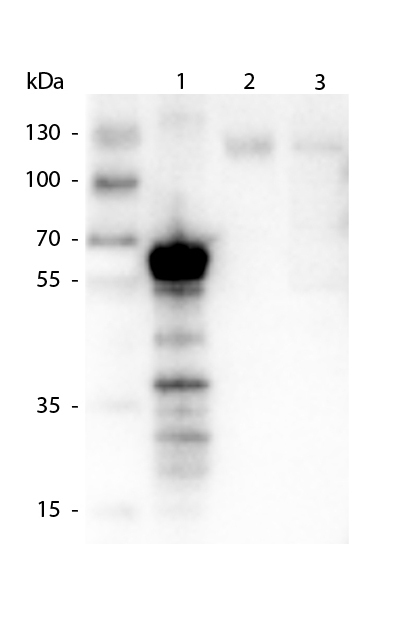
- Western Blot of Mouse anti-AKT3 antibody. Lane 1: Control. Lane 2: Rapa. Lane 3: T50. Lane 4: T250. Lane 5: Control. Lane 6: Rapa. Lane 7: T50. Lane 8: T250. Lane 9: AKT3 null. Load: 35 µg per lane. Primary antibody: AKT-3 antibody at 1:1000 for overnight at 4°C. Secondary antibody: Anti mouse secondary antibody at 1:20,000 for 1 h at RT. Block: 5% BLOTTO overnight at 4°C. Predicted/Observed size: 56 kDa for AKT3.
Enhanced validation
- Submitted by
- LSBio (provider)
- Enhanced method
- Genetic validation
- Main image
- Experimental details
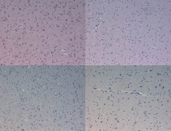
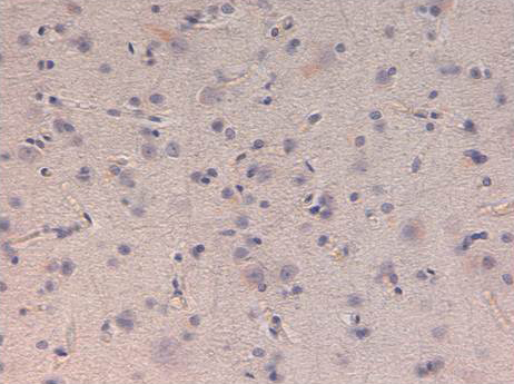
- Immunohistochemistry of Mouse Anti-AKT3 antibody. Tissue: human prostate carcinoma. A) AKT-3 antibody produced using CELLine, B) AKT-3 antibody produced using roller bottle. Fixation: formalin fixed paraffin embedded. Antigen retrieval: not required. Primary antibody: AKT-3 antibody at 10 µg/mL for 1 h at RT. Secondary antibody: Peroxidase mouse secondary antibody at 1:10,000 for 1 h at RT. Localization: AKT3 is nuclear and occasionally cytoplasmic. Staining: AKT3 as precipitated brown signal with hematoxylin purple nuclear counterstain.

- Submitted by
- LSBio (provider)
- Enhanced method
- Genetic validation
- Main image

- Experimental details
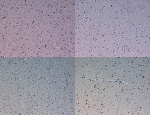
- Immunohistochemistry of Mouse Anti-AKT3 antibody. Tissue: human prostate carcinoma. A) AKT-3 antibody produced using CELLine, B) AKT-3 antibody produced using roller bottle. Fixation: formalin fixed paraffin embedded. Antigen retrieval: not required. Primary antibody: AKT-3 antibody at 10 µg/mL for 1 h at RT. Secondary antibody: Peroxidase mouse secondary antibody at 1:10,000 for 1 h at RT. Localization: AKT3 is nuclear and occasionally cytoplasmic. Staining: AKT3 as precipitated brown signal with hematoxylin purple nuclear counterstain.
- Submitted by
- LSBio (provider)
- Enhanced method
- Genetic validation
- Main image

- Experimental details
- Western Blot of Mouse Anti-AKT3 antibody. Lane 1: C2C12. Lane 2: MEF#1. Lane 3: MEF#2. Lane 4: A549. Lane 5: Calu-1. Lane 6: PC3. Lane 7: HepG2. Lane 8: Jurkat. Lane 9: SKOV3. Lane 10: 293T. Load: 35 µg per lane. Primary antibody: AKT-3 antibody at 1:1000 for overnight at 4°C. Secondary antibody: Anti mouse secondary antibody at 1:20,000 for 1 h at RT. Block: 5% BLOTTO overnight at 4°C. Predicted/Observed size: 56 kDa for AKT3.

- Submitted by
- LSBio (provider)
- Main image
- Experimental details
- Western Blot of Mouse Anti-AKT3 antibody. Lane 1: C2C12. Lane 2: MEF#1. Lane 3: MEF#2. Lane 4: A549. Lane 5: Calu-1. Lane 6: PC3. Lane 7: HepG2. Lane 8: Jurkat. Lane 9: SKOV3. Lane 10: 293T. Load: 35 µg per lane. Primary antibody: AKT-3 antibody at 1:1000 for overnight at 4°C. Secondary antibody: Anti mouse secondary antibody at 1:20,000 for 1 h at RT. Block: 5% BLOTTO overnight at 4°C. Predicted/Observed size: 56 kDa for AKT3.
- Submitted by
- LSBio (provider)
- Main image
- Experimental details
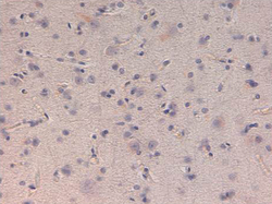
- Immunohistochemistry of Mouse Anti-AKT3 antibody. Tissue: human prostate carcinoma. A) AKT-3 antibody produced using CELLine, B) AKT-3 antibody produced using roller bottle. Fixation: formalin fixed paraffin embedded. Antigen retrieval: not required. Primary antibody: AKT-3 antibody at 10 µg/mL for 1 h at RT. Secondary antibody: Peroxidase mouse secondary antibody at 1:10,000 for 1 h at RT. Localization: AKT3 is nuclear and occasionally cytoplasmic. Staining: AKT3 as precipitated brown signal with hematoxylin purple nuclear counterstain.
