 Explore
 Validate
 Learn
Learn Western blot
 Immunocytochemistry
Antibody data
- Antibody Data
- Antigen structure
- References [2]
- Comments [0]
- Validations
- Immunocytochemistry [2]
- Immunoprecipitation [1]
- Other assay [1]
Submit
Validation data
Reference
Comment
Report error
- Product number
- PA1-643 - Provider product page

- Provider
- Invitrogen Antibodies
- Product name
- Kinesin 5B Polyclonal Antibody
- Antibody type
- Polyclonal
- Antigen
- Synthetic peptide
- Description
- PA1-643 detects kinesin 5B from mouse and human samples. This antibody is specific for kinesin 5B and does not detect other kinesin isotypes. PA1-643 has been successfully used in Western blot and immunofluorescent procedures. By Western blot, this antibody detects an ~110 kDa protein representing kinesin 5B protein. The PA1-643 immunizing peptide corresponds to amino acid residues 376-396 from human kinesin 5B. This sequence is 95% and 90% conserved for mouse and rat, respectively. This peptide (Cat. # PEP-198) is available for use in neutralization and control experiments.
- Reactivity
- Human, Mouse, Rat
- Host
- Rabbit
- Isotype
- IgG
- Vial size
- 100 μg
- Concentration
- 1 mg/mL
- Storage
- -20°C, Avoid Freeze/Thaw Cycles
Submitted references Daxx functions as a scaffold of a protein assembly constituted by GLUT4, JNK1 and KIF5B.
Conventional kinesin holoenzymes are composed of heavy and light chain homodimers.
Lalioti VS, Vergarajauregui S, Tsuchiya Y, Hernandez-Tiedra S, Sandoval IV
Journal of cellular physiology 2009 Feb;218(2):416-26
Journal of cellular physiology 2009 Feb;218(2):416-26
Conventional kinesin holoenzymes are composed of heavy and light chain homodimers.
DeBoer SR, You Y, Szodorai A, Kaminska A, Pigino G, Nwabuisi E, Wang B, Estrada-Hernandez T, Kins S, Brady ST, Morfini G
Biochemistry 2008 Apr 15;47(15):4535-43
Biochemistry 2008 Apr 15;47(15):4535-43
No comments: Submit comment
Supportive validation
- Submitted by
- Invitrogen Antibodies (provider)
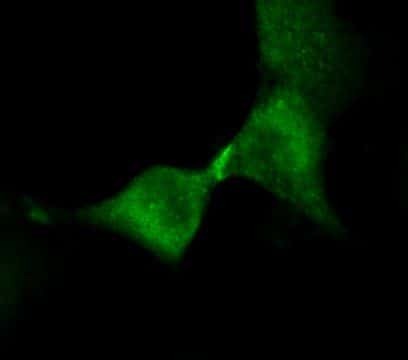
- Main image

- Experimental details
- Immunofluorescent analysis of Kinesin 5B (green) in HeLa cells. Cells fixed in 3.2% paraformaldehyde and permeabilized with 0.2% Triton X-100 for 30 minutes at 37°C were blocked with 3% BSA in PBS for 30 minutes at room temperature. Cells were probed with a Kinesin 5B polyclonal antibody (Product # PA1-643) at a dilution of 1:250 overnight at 4°C, washed with PBS containing 0.2% Triton X-100, and incubated with a fluorescently-conjugated goat anti-rabbit IgG secondary antibody at a dilution of 1:2000 for 1 hour at room temperature. Images were taken on wide-field fluorescent microscope at 63X magnification. Data courtesy of the Innovators Program.
- Submitted by
- Invitrogen Antibodies (provider)
- Main image

- Experimental details
- Immunofluorescent analysis of Kinesin 5B (green) in HeLa cells. Cells fixed in 3.2% paraformaldehyde and permeabilized with 0.2% Triton X-100 for 30 minutes at 37°C were blocked with 3% BSA in PBS for 30 minutes at room temperature. Cells were probed with a Kinesin 5B polyclonal antibody (Product # PA1-643) at a dilution of 1:250 overnight at 4°C, washed with PBS containing 0.2% Triton X-100, and incubated with a fluorescently-conjugated goat anti-rabbit IgG secondary antibody at a dilution of 1:2000 for 1 hour at room temperature. Images were taken on wide-field fluorescent microscope at 63X magnification. Data courtesy of the Innovators Program.

Supportive validation
- Submitted by
- Invitrogen Antibodies (provider)
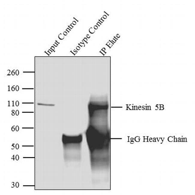
- Main image

- Experimental details
- Kinesin 5B was immunoprecipitated using 5 µg of the Kinesin 5B Rabbit Polyclonal Antibody (Product # PA1-643) from lysate of Mouse Brain (Lane 3) using the Dynabeads® Protein A Immunoprecipitation Kit (Product # 10006D). Normal Rabbit IgG was used as a Isotype control (Lane 2). 10 % input represents the cell extract used for immunoprecipitation (Lane 1). Western blot analysis was performed using Kinesin 5B Rabbit Polyclonal Antibody (Product # PA1-643) and Goat anti-Rabbit IgG (Heavy Chain) Superclonal™ Secondary Antibody, HRP conjugate (Product # A27036, 0.4 µg/mL, 1:2500 dilution). Chemiluminescent detection was performed using Pierce™ ECL Western Blotting Substrate (Product # 32106).
Supportive validation
- Submitted by
- Invitrogen Antibodies (provider)
- Main image

- Experimental details
- Kinesin 5B was immunoprecipitated using 5 æg of the Kinesin 5B Rabbit Polyclonal Antibody (Product # PA1-643) from lysate of Mouse Brain (Lane 3) using the Dynabeads® Protein A Immunoprecipitation Kit (Product # 10006D). Normal Rabbit IgG was used as a Isotype control (Lane 2). 10 % input represents the cell extract used for immunoprecipitation (Lane 1). Western blot analysis was performed using Kinesin 5B Rabbit Polyclonal Antibody (Product # PA1-643) and Goat anti-Rabbit IgG (H+L) Superclonal™ Secondary Antibody, HRP conjugate (Product # A27036, 0.4 æg/mL, 1:2500 dilution). Chemiluminescent detection was performed using Pierce™ ECL Western Blotting Substrate (Product # 32106).
