Explore
 Validate
 Learn
Learn Western blot
 Immunocytochemistry
Antibody data
- Antibody Data
- Antigen structure
- References [0]
- Comments [0]
- Validations
- Immunocytochemistry [2]
- Immunoprecipitation [1]
- Immunohistochemistry [4]
- Other assay [1]
Submit
Validation data
Reference
Comment
Report error
- Product number
- PA5-29948 - Provider product page

- Provider
- Invitrogen Antibodies
- Product name
- SFPQ Polyclonal Antibody
- Antibody type
- Polyclonal
- Antigen
- Recombinant full-length protein
- Description
- Recommended positive controls: 293T, A431, HeLa, HepG2, Neuro 2A, C8D30, NIH-3T3, Raw264.7, C2C12. Predicted reactivity: Mouse (100%), Rat (100%), Zebrafish (85%), Chimpanzee (100%), Bovine (100%). Store product as a concentrated solution. Centrifuge briefly prior to opening the vial.
- Reactivity
- Human, Mouse
- Host
- Rabbit
- Isotype
- IgG
- Vial size
- 100 μL
- Concentration
- 0.78 mg/mL
- Storage
- Store at 4°C short term. For long term storage, store at -20°C, avoiding freeze/thaw cycles.
No comments: Submit comment
Supportive validation
- Submitted by
- Invitrogen Antibodies (provider)
- Main image

- Experimental details
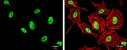
- Immunocytochemistry-Immunofluorescence analysis of SFPQ was performed in HeLa cells fixed in 4% paraformaldehyde at RT for 15 min. Green: SFPQ Polyclonal Antibody (Product # PA5-29948) diluted at 1:500. Red: phalloidin, a cytoskeleton marker. Scale bar = 10 µm.

- Submitted by
- Invitrogen Antibodies (provider)
- Main image
- Experimental details
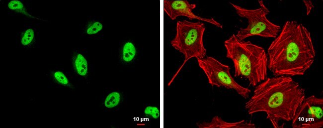
- Immunocytochemistry-Immunofluorescence analysis of SFPQ was performed in HeLa cells fixed in 4% paraformaldehyde at RT for 15 min. Green: SFPQ Polyclonal Antibody (Product # PA5-29948) diluted at 1:500. Red: phalloidin, a cytoskeleton marker. Scale bar = 10 µm.
Supportive validation
- Submitted by
- Invitrogen Antibodies (provider)
- Main image
- Experimental details
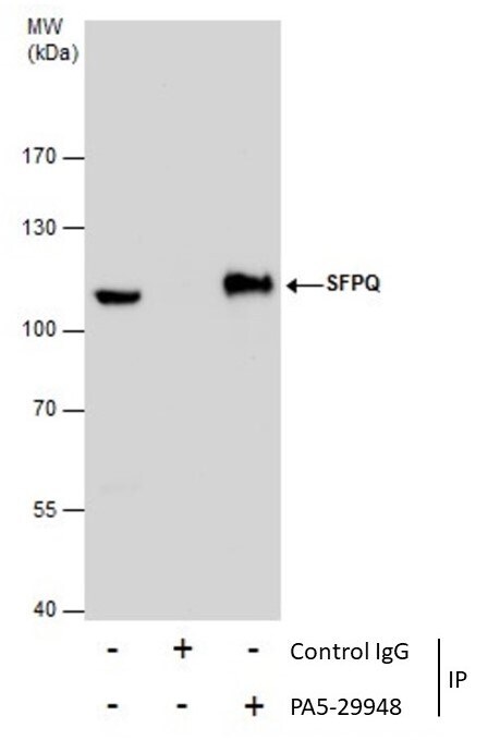
- Immunoprecipitation of SFPQ was performed in 293T whole cell extracts using 5 µg of SFPQ Polyclonal Antibody (Product # PA5-29948). Samples were transferred to a membrane and probed with SFPQ Polyclonal Antibody as a primary antibody and an HRP-conjugated anti-Rabbit IgG was used as a secondary antibody.

Supportive validation
- Submitted by
- Invitrogen Antibodies (provider)
- Main image
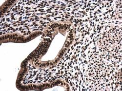
- Experimental details
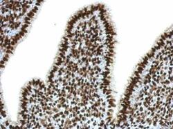
- Immunohistochemical analysis of paraffin-embedded mouse uterus, using SFPQ (Product # PA5-29948) antibody at 1:500 dilution. Antigen Retrieval: EDTA based buffer, pH 8.0, 15 min.

- Submitted by
- Invitrogen Antibodies (provider)
- Main image

- Experimental details
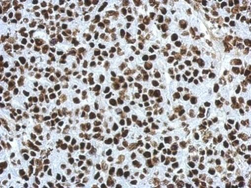
- Immunohistochemical analysis of paraffin-embedded Hela xenograft, using SFPQ (Product # PA5-29948) antibody at 1:500 dilution. Antigen Retrieval: EDTA based buffer, pH 8.0, 15 min.
- Submitted by
- Invitrogen Antibodies (provider)
- Main image
- Experimental details
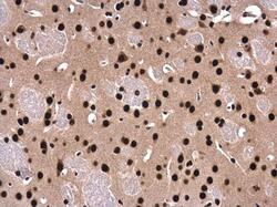
- Immunohistochemistry (Paraffin) analysis of SFPQ was performed in paraffin-embedded mouse fore brain tissue using SFPQ Polyclonal Antibody (Product # PA5-29948) at a dilution of 1:500.
- Submitted by
- Invitrogen Antibodies (provider)
- Main image
- Experimental details
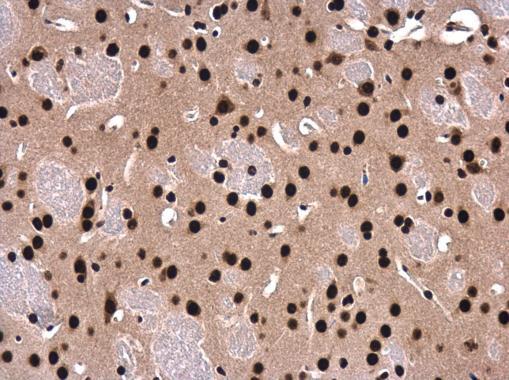
- Immunohistochemistry (Paraffin) analysis of SFPQ was performed in paraffin-embedded mouse cervix tissue using SFPQ Polyclonal Antibody (Product # PA5-29948) at a dilution of 1:500.

Supportive validation
- Submitted by
- Invitrogen Antibodies (provider)
- Main image

- Experimental details
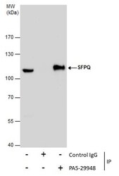
- Immunoprecipitation of SFPQ was performed in 293T whole cell extracts using 5 µg of SFPQ Polyclonal Antibody (Product # PA5-29948). Samples were transferred to a membrane and probed with SFPQ Polyclonal Antibody as a primary antibody and an HRP-conjugated anti-Rabbit IgG was used as a secondary antibody.