 Explore
 Validate
 Learn
Learn Flow cytometry
Antibody data
- Antibody Data
- Antigen structure
- References [2]
- Comments [0]
- Validations
- Flow cytometry [1]
Submit
Validation data
Reference
Comment
Report error
- Product number
- FAB9181G - Provider product page

- Provider
- Novus Biologicals
- Product name
- Mouse Monoclonal MMP-14/MT1-MMP Antibody
- Antibody type
- Monoclonal
- Description
- Protein A or G purified from hybridoma culture supernatant. Detects human MMP-14/MT1-MMP in direct ELISAs and Western blots. Does not detect E. coli-expressed recombinant human MMP-14 catalytic domain (aa 112-284).
- Reactivity
- Human
- Host
- Mouse
- Conjugate
- Green dye
- Isotype
- IgG
- Vial size
- 100 Tests
- Storage
- Protect from light. Do not freeze. 12 months from date of receipt, 2 to 8 degreesC as supplied.
Submitted references A PKA/cdc42 Signaling Axis Restricts Angiogenic Sprouting by Regulating Podosome Rosette Biogenesis and Matrix Remodeling.
NEDD9/Arf6-dependent endocytic trafficking of matrix metalloproteinase 14: a novel mechanism for blocking mesenchymal cell invasion and metastasis of breast cancer.
MacKeil JL, Brzezinska P, Burke-Kleinman J, Craig AW, Nicol CJB, Maurice DH
Scientific reports 2019 Feb 20;9(1):2385
Scientific reports 2019 Feb 20;9(1):2385
NEDD9/Arf6-dependent endocytic trafficking of matrix metalloproteinase 14: a novel mechanism for blocking mesenchymal cell invasion and metastasis of breast cancer.
Loskutov YV, Kozyulina PY, Kozyreva VK, Ice RJ, Jones BC, Roston TJ, Smolkin MB, Ivanov AV, Wysolmerski RB, Pugacheva EN
Oncogene 2015 Jul;34(28):3662-75
Oncogene 2015 Jul;34(28):3662-75
No comments: Submit comment
Supportive validation
- Submitted by
- Novus Biologicals (provider)
- Main image
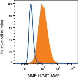
- Experimental details
- Detection of MMP-14/MT1-MMP in MDA-MB-231 Human Cell Line by Flow Cytometry. MDA-MB-231 human breast cancer cell line was stained with Mouse Anti-Human MMP-14/MT1-MMP Alexa Fluor® 488-conjugated Monoclonal Antibody (Catalog # FAB9181G, filled histogram) or isotype control antibody (Catalog # IC0041G, open histogram). View our protocol for Staining Membrane-associated Proteins.
