Explore
 Validate
 Learn
Learn Western blot
 Immunocytochemistry
Antibody data
- Antibody Data
- Antigen structure
- References [4]
- Comments [0]
- Validations
- Immunocytochemistry [2]
- Immunohistochemistry [1]
- Flow cytometry [2]
- Other assay [1]
Submit
Validation data
Reference
Comment
Report error
- Product number
- PA5-13183 - Provider product page

- Provider
- Invitrogen Antibodies
- Product name
- MMP14 Polyclonal Antibody
- Antibody type
- Polyclonal
- Antigen
- Synthetic peptide
- Description
- This antibody is predicted to react with bovine, mouse, porcine and rat based on sequence homology.
- Reactivity
- Human, Mouse, Rat
- Host
- Rabbit
- Isotype
- IgG
- Vial size
- 400 μL
- Concentration
- 2 mg/mL
- Storage
- Store at 4°C short term. For long term storage, store at -20°C, avoiding freeze/thaw cycles.
Submitted references Mesenchymal stromal cell-derived septoclasts resorb cartilage during developmental ossification and fracture healing.
The Effect of Neddylation Inhibition on Inflammation-Induced MMP9 Gene Expression in Esophageal Squamous Cell Carcinoma.
TNF-α promotes breast cancer cell migration and enhances the concentration of membrane-associated proteases in lipid rafts.
Serous and mucinous ovarian tumors express different profiles of MMP-2, -7, -9, MT1-MMP, and TIMP-1 and -2.
Sivaraj KK, Majev PG, Jeong HW, Dharmalingam B, Zeuschner D, Schröder S, Bixel MG, Timmen M, Stange R, Adams RH
Nature communications 2022 Jan 28;13(1):571
Nature communications 2022 Jan 28;13(1):571
The Effect of Neddylation Inhibition on Inflammation-Induced MMP9 Gene Expression in Esophageal Squamous Cell Carcinoma.
Hryniewicz-Jankowska A, Wierzbicki J, Tabola R, Stach K, Sossey-Alaoui K, Augoff K
International journal of molecular sciences 2021 Feb 9;22(4)
International journal of molecular sciences 2021 Feb 9;22(4)
TNF-α promotes breast cancer cell migration and enhances the concentration of membrane-associated proteases in lipid rafts.
Wolczyk D, Zaremba-Czogalla M, Hryniewicz-Jankowska A, Tabola R, Grabowski K, Sikorski AF, Augoff K
Cellular oncology (Dordrecht) 2016 Aug;39(4):353-63
Cellular oncology (Dordrecht) 2016 Aug;39(4):353-63
Serous and mucinous ovarian tumors express different profiles of MMP-2, -7, -9, MT1-MMP, and TIMP-1 and -2.
Brun JL, Cortez A, Commo F, Uzan S, Rouzier R, Daraï E
International journal of oncology 2008 Dec;33(6):1239-46
International journal of oncology 2008 Dec;33(6):1239-46
No comments: Submit comment
Supportive validation
- Submitted by
- Invitrogen Antibodies (provider)
- Main image
- Experimental details
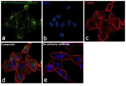
- Immunofluorescence analysis of MMP14 was performed using 70% confluent log phase MDA-MB-231 cells. The cells were fixed with 4% paraformaldehyde for 10 minutes, permeabilized with 0.1% Triton™ X-100 for 15 minutes, and blocked with 1% BSA for 1 hour at room temperature. The cells were labeled with MMP14 Polyclonal Antibody (Product # PA5-13183) at 1:100 dilution in 0.1% BSA, incubated at 4 degree Celsius overnight and then labeled with Goat anti-Rabbit IgG (H+L) Superclonal™ Secondary Antibody, Alexa Fluor® 488 conjugate (Product # A27034) for 45 minutes at room temperature (Panel a: green). Nuclei (Panel b: blue) were stained with ProLong™ Diamond Antifade Mountant with DAPI (Product # P36962). F-actin (Panel c: red) was stained with Rhodamine Phalloidin (Product # R415, 1:300). Panel d represents the merged image showing golgi and cytoplasmic localization. Panel e represents control cells with no primary antibody to assess background. The images were captured at 60X magnification.

- Submitted by
- Invitrogen Antibodies (provider)
- Main image

- Experimental details
- Immunofluorescence analysis of MMP14 was performed using 70% confluent log phase MDA-MB-231 cells. The cells were fixed with 4% paraformaldehyde for 10 minutes, permeabilized with 0.1% Triton™ X-100 for 15 minutes, and blocked with 1% BSA for 1 hour at room temperature. The cells were labeled with MMP14 Polyclonal Antibody (Product # PA5-13183) at 1:100 dilution in 0.1% BSA, incubated at 4 degree Celsius overnight and then labeled with Goat anti-Rabbit IgG (Heavy Chain) Superclonal™ Secondary Antibody, Alexa Fluor® 488 conjugate (Product # A27034) for 45 minutes at room temperature (Panel a: green). Nuclei (Panel b: blue) were stained with ProLong™ Diamond Antifade Mountant with DAPI (Product # P36962). F-actin (Panel c: red) was stained with Rhodamine Phalloidin (Product # R415, 1:300). Panel d represents the merged image showing golgi and cytoplasmic localization. Panel e represents control cells with no primary antibody to assess background. The images were captured at 60X magnification.

Supportive validation
- Submitted by
- Invitrogen Antibodies (provider)
- Main image
- Experimental details
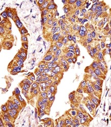
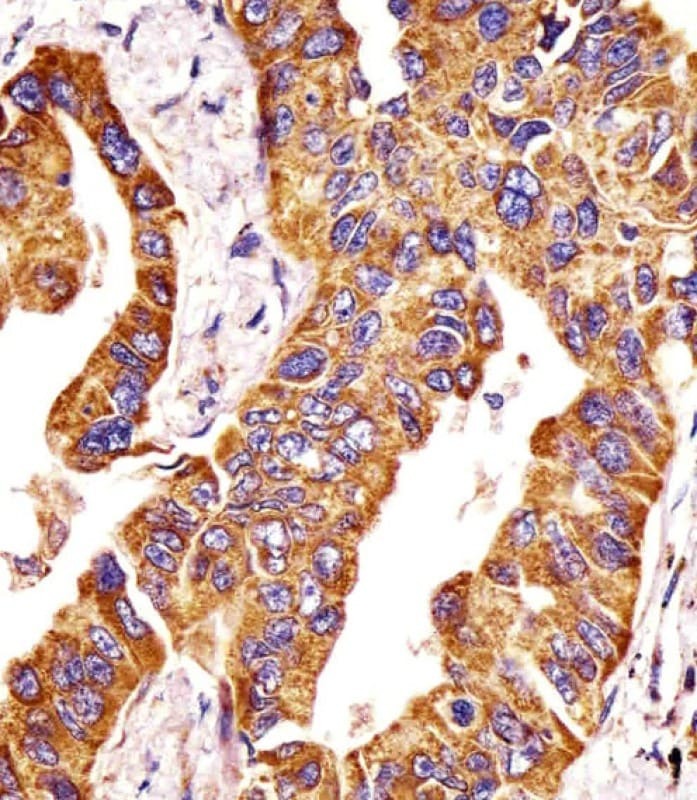
- Immunohistochemistry analysis of MMP14 in paraformaldehyde-fixed, paraffin-embedded human lung adenocarcinoma tissue sections. Samples were incubated with MMP14 polyclonal antibody (Product # PA5-13183) using a dilution of 1:25 for 1 hours at 37°C followed by an undiluted biotinylated goat polyvalent antibody. Tissue was fixed with formaldehyde and blocked with 3% BSA for 0.5 hour at room temperature; antigen retrieval was by heat mediation with a citrate buffer (pH 6).
Supportive validation
- Submitted by
- Invitrogen Antibodies (provider)
- Main image
- Experimental details
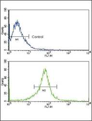
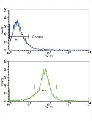
- Flow cytometry analysis of MCF-7 cells using a MMP14 polyclonal antibody (Product # PA5-13183) (bottom), compared to a negative control cell (top) at a dilution of 1:10-50, followed by a FITC-conjugated goat anti-rabbit antibody
- Submitted by
- Invitrogen Antibodies (provider)
- Main image
- Experimental details
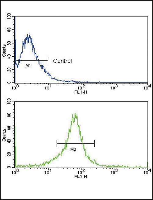
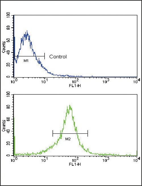
- Flow cytometry of MMP14 in MCF-7 cells (bottom histogram). Samples were incubated with MMP14 polyclonal antibody (Product # PA5-13183) followed by FITC-conjugated goat-anti-rabbit secondary antibody. Negative control (top histogram).
Supportive validation
- Submitted by
- Invitrogen Antibodies (provider)
- Main image
- Experimental details
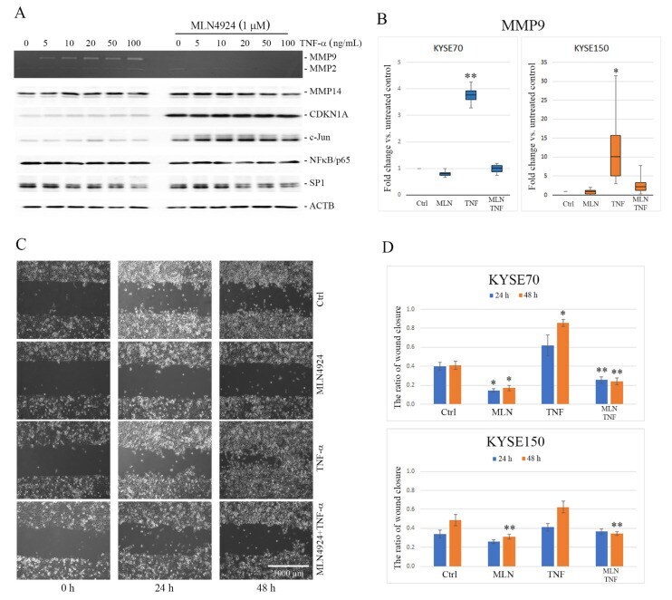
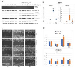
- Figure 1 Effect of tumor necrosis factor-alpha (TNF-alpha) and MLN4924 on matrix metalloproteinase 9 (MMP9) expression and esophageal squamous cell carcinoma (ESCC) cell migration. ( A ) After overnight serum starvation, KYSE150 cells were pretreated or not with 1 muM MLN4924 for 30 min and then stimulated with TNF-alpha at the indicated concentration for 24 h. Activity of MMP9 and MMP2 was analyzed by gelatin zymography in the conditioned media. The protein levels of membrane type I-matrix metalloproteinase (MT1-MMP), cyclin dependent kinase inhibitor 1A (CDKN1A/p21), c-Jun, nuclear factor kappa B (NFkappaB) and SP1 were determined by Western blotting. Fold change calculated as the ratio of relative levels of proteins normalized to beta-actin (ACTB) between the cells treated with TNF-alpha in combination with MLN4924 and the cells treated with TNF-alpha alone is shown in Figure S4 . Values shown are means +- SEM: whiskers: min-max. ( B ) MMP9 messenger RNA (mRNA) expression was analyzed by reverse transcription quantitative real-time polymerase chain reaction (RT-qPCR) in both KYSE150 and KYSE70 cells treated for 24 h with TNF-alpha (30 ng/mL) and MLN4924 (1 µM) alone or in combination. Results are presented as fold change of MMP9 gene in the treated cells relative to the untreated controls normalized to the expression of the reference gene encoding glyceraldehyde 3-phosphate dehydrogenase ( GAPDH ). ( C ) Representative images from wound healing assay showing the chan