Explore
 Validate
 Learn
Learn Western blot
Antibody data
- Antibody Data
- Antigen structure
- References [0]
- Comments [0]
- Validations
- Western blot [5]
- Immunohistochemistry [1]
Submit
Validation data
Reference
Comment
Report error
- Product number
- AP8087a - Provider product page

- Provider
- Abcepta
- Proper citation
- Abgent Cat#AP8087a, RRID:AB_2161841
- Product name
- PACSIN1 Antibody (N-term)
- Antibody type
- Polyclonal
- Antigen
- Synthetic peptide
- Description
- Purified Rabbit Polyclonal Antibody (Pab)
- Reactivity
- Human, Mouse
- Host
- Rabbit
- Isotype
- IgG
- Vial size
- 400 µl
- Concentration
- 2 mg/ml
- Storage
- Maintain refrigerated at 2-8°C for up to 6 months. For long term storage store at -20°C in small aliquots to prevent freeze-thaw cycles.
No comments: Submit comment
Supportive validation
- Submitted by
- Abcepta (provider)
- Main image
- Experimental details
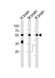
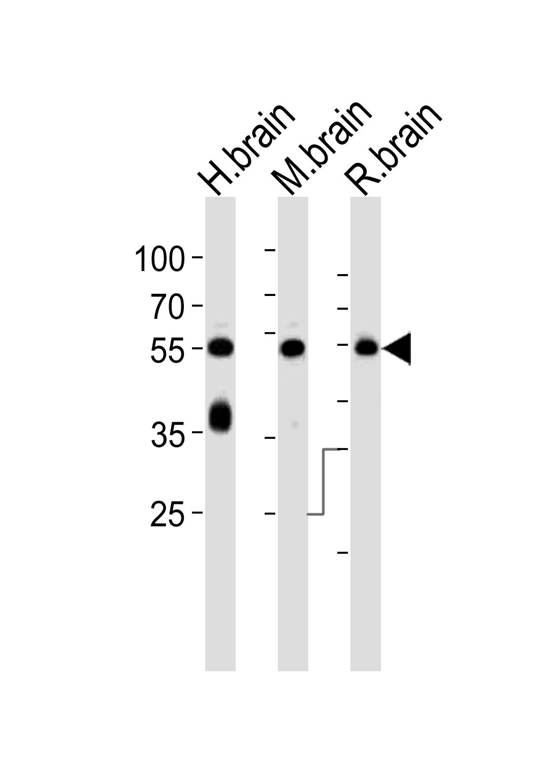
- "Western blot analysis of lysates from human brain, mouse brain and rat brain tissue lysate (from left to right), using PACSIN1 Antibody (G23)(Cat. #AP8087a). AP8087a was diluted at 1:1000 at each lane. A goat anti-rabbit IgG H&L(HRP) at 1:10000 dilution was used as the secondary antibody. Lysates at 35ug per lane."
- Primary Ab dilution
- 1:1000
- Submitted by
- Abcepta (provider)
- Main image
- Experimental details
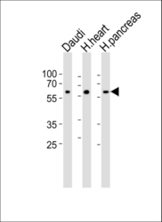
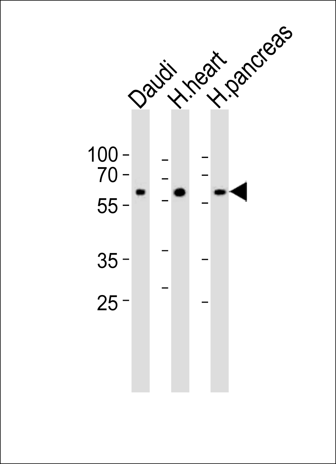
- "Western blot analysis of lysates from Daudi cell line, human heart and pancreas tissue lysate(from left to right), using PACSIN1 Antibody (G23)(Cat. #AP8087a). AP8087a was diluted at 1:1000 at each lane. A goat anti-rabbit IgG H&L(HRP) at 1:10000 dilution was used as the secondary antibody. Lysates at 35ug per lane."
- Primary Ab dilution
- 1:1000
- Submitted by
- Abcepta (provider)
- Main image
- Experimental details
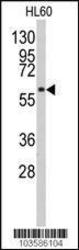
- Western blot analysis of anti-PACSIN1 Pab (Cat. #AP8087a) in HL-60 cell line lysate. PACSIN1 (arrow) was detected using purified Pab. Secondary HRP-anti-rabbit was used for signal visualization with chemiluminescence.
- Primary Ab dilution
- 1:1000

- Submitted by
- Abcepta (provider)
- Main image
- Experimental details
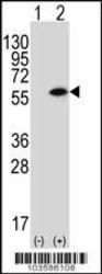
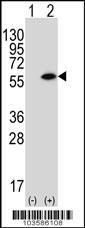
- Western blot analysis of PACSIN1 (arrow) using rabbit polyclonal PACSIN1 Antibody (G23) (Cat. #AP8087a). 293 cell lysates (2 ug/lane) either nontransfected (Lane 1) or transiently transfected (Lane 2) with the PACSIN1 gene.
- Primary Ab dilution
- 1:1000
- Submitted by
- Abcepta (provider)
- Main image
- Experimental details
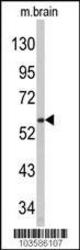
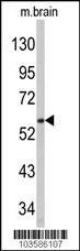
- Western blot analysis of anti-PACSIN1 Pab (Cat. #AP8087a) in mouse brain tissue lysate . PACSIN1 (arrow) was detected using purified Pab. Secondary HRP-anti-rabbit was used for signal visualization with chemiluminescence.
- Primary Ab dilution
- 1:1000
Supportive validation
- Submitted by
- Abcepta (provider)
- Main image
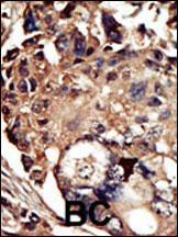
- Experimental details
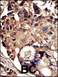
- "Formalin-fixed and paraffin-embedded human cancer tissue reacted with the primary antibody, which was peroxidase-conjugated to the secondary antibody, followed by DAB staining. This data demonstrates the use of this antibody for immunohistochemistry; clinical relevance has not been evaluated. BC = breast carcinoma; HC = hepatocarcinoma."
- Primary Ab dilution
- 1:50~100