Explore
 Validate
 Learn
Learn Western blot
 Immunohistochemistry
Antibody data
- Antibody Data
- Antigen structure
- References [2]
- Comments [0]
- Validations
- Immunohistochemistry [1]
Submit
Validation data
Reference
Comment
Report error
- Product number
- HPA028852 - Provider product page

- Provider
- Atlas Antibodies
- Proper citation
- Atlas Antibodies Cat#HPA028852, RRID:AB_10602812
- Product name
- Anti-PACSIN1
- Antibody type
- Polyclonal
- Description
- Polyclonal Antibody against Human PACSIN1, Gene description: protein kinase C and casein kinase substrate in neurons 1, Alternative Gene Names: SDPI, Validated applications: IHC, WB, Uniprot ID: Q9BY11, Storage: Store at +4°C for short term storage. Long time storage is recommended at -20°C.
- Reactivity
- Human
- Host
- Rabbit
- Conjugate
- Unconjugated
- Isotype
- IgG
- Vial size
- 100 µl
- Concentration
- 0.05 mg/ml
- Storage
- Store at +4°C for short term storage. Long time storage is recommended at -20°C.
- Handling
- The antibody solution should be gently mixed before use.
Submitted references PACSIN1 promotes immunosuppression in gastric cancer by degrading MHC-I
Immunofluorescence and fluorescent-protein tagging show high correlation for protein localization in mammalian cells
Liu Z, Li X, Muhammad A, Sun Q, Zhang Q, Wang Y, Wang Y, Ren J, Wang D
Acta Biochimica et Biophysica Sinica 2024;56(10):1473-1482
Acta Biochimica et Biophysica Sinica 2024;56(10):1473-1482
Immunofluorescence and fluorescent-protein tagging show high correlation for protein localization in mammalian cells
Stadler C, Rexhepaj E, Singan V, Murphy R, Pepperkok R, Uhlén M, Simpson J, Lundberg E
Nature Methods 2013;10(4):315-323
Nature Methods 2013;10(4):315-323
No comments: Submit comment
Supportive validation
- Submitted by
- Atlas Antibodies (provider)
- Enhanced method
- Orthogonal validation
- Main image

- Experimental details
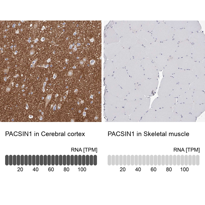
- Immunohistochemistry analysis in human cerebral cortex and skeletal muscle tissues using Anti-PACSIN1 antibody. Corresponding PACSIN1 RNA-seq data are presented for the same tissues.
- Sample type
- Human
- Protocol
- Protocol