 Explore
 Validate
 Learn
Learn Western blot
 Immunocytochemistry
 Immunohistochemistry
Antibody data
- Antibody Data
- Antigen structure
- References [1]
- Comments [0]
- Validations
- Immunocytochemistry [4]
Submit
Validation data
Reference
Comment
Report error
- Product number
- MA1-23328 - Provider product page

- Provider
- Invitrogen Antibodies
- Product name
- XPC Monoclonal Antibody (3.26)
- Antibody type
- Monoclonal
- Antigen
- Purifed from natural sources
- Description
- Recommended positive controls: HeLa whole cell extract. Store product as a concentrated solution. Centrifuge briefly prior to opening the vial.
- Reactivity
- Human, Mouse
- Host
- Mouse
- Isotype
- IgG
- Antibody clone number
- 3.26
- Vial size
- 100 μL
- Concentration
- 1.15 mg/mL
- Storage
- Store at 4°C short term. For long term storage, store at -20°C, avoiding freeze/thaw cycles.
Submitted references CRISPR-Pass: Gene Rescue of Nonsense Mutations Using Adenine Base Editors.
Lee C, Hyun Jo D, Hwang GH, Yu J, Kim JH, Park SE, Kim JS, Kim JH, Bae S
Molecular therapy : the journal of the American Society of Gene Therapy 2019 Aug 7;27(8):1364-1371
Molecular therapy : the journal of the American Society of Gene Therapy 2019 Aug 7;27(8):1364-1371
No comments: Submit comment
Supportive validation
- Submitted by
- Invitrogen Antibodies (provider)
- Main image
- Experimental details
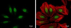
- XPC Monoclonal Antibody (3.26) detects XPC protein at nucleus by immunofluorescent analysis. Sample: HeLa cells were fixed in 4% paraformaldehyde at RT for 15 min. Green: XPC protein stained by XPC Monoclonal Antibody (3.26) (Product # MA1-23328) diluted at 1:100. Red: Phalloidin, a cytoskeleton marker, diluted at 1:200. Scale bar = 10 µm.

- Submitted by
- Invitrogen Antibodies (provider)
- Main image

- Experimental details
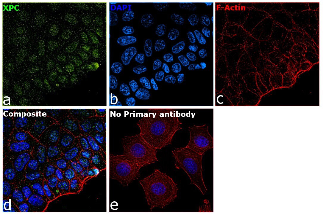
- Immunofluorescence analysis of XPC was performed using 70% confluent log phase MCF7 cells. The cells were fixed with 4% paraformaldehyde for 10 minutes, permeabilized with 0.1% Triton™ X-100 for 15 minutes, and blocked with 2% BSA for 1 hour at room temperature. The cells were labeled with XPC Monoclonal Antibody (3.26) (Product # MA1-23328) at 5 µg/mL in 0.1% BSA, incubated at 4 degree Celsius overnight and then with Goat anti-Mouse IgG (H+L), Superclonal™ Recombinant Secondary Antibody, Alexa Fluor 488 conjugate (Product # A28175) at a dilution of 1:2000 for 45 minutes at room temperature (Panel a: green). Nuclei (Panel b: blue) were stained with SlowFade® Gold Antifade Mountant with DAPI (Product # S36938). F-actin (Panel c: red) was stained with Rhodamine Phalloidin (Product # R415, 1:300). Panel d represents the merged image showing staining in nucleus. Panel e represents control cells with no primary antibody to assess background. The images were captured at 60X magnification.

- Submitted by
- Invitrogen Antibodies (provider)
- Main image

- Experimental details
- Immunofluorescence analysis of XPC was performed using 70% confluent log phase MCF7 cells. The cells were fixed with 4% paraformaldehyde for 10 minutes, permeabilized with 0.1% Triton™ X-100 for 15 minutes, and blocked with 2% BSA for 1 hour at room temperature. The cells were labeled with XPC Monoclonal Antibody (3.26) (Product # MA1-23328) at 5 µg/mL in 0.1% BSA, incubated at 4 degree Celsius overnight and then with Goat anti-Mouse IgG (H+L), Superclonal™ Recombinant Secondary Antibody, Alexa Fluor 488 conjugate (Product # A28175) at a dilution of 1:2000 for 45 minutes at room temperature (Panel a: green). Nuclei (Panel b: blue) were stained with SlowFade® Gold Antifade Mountant with DAPI (Product # S36938). F-actin (Panel c: red) was stained with Rhodamine Phalloidin (Product # R415, 1:300). Panel d represents the merged image showing staining in nucleus. Panel e represents control cells with no primary antibody to assess background. The images were captured at 60X magnification.
- Submitted by
- Invitrogen Antibodies (provider)
- Main image

- Experimental details
- XPC Monoclonal Antibody (3.26) detects XPC protein at nucleus by immunofluorescent analysis. Sample: HeLa cells were fixed in 4% paraformaldehyde at RT for 15 min. Green: XPC protein stained by XPC Monoclonal Antibody (3.26) (Product # MA1-23328) diluted at 1:100. Red: Phalloidin, a cytoskeleton marker, diluted at 1:200. Scale bar = 10 µm.
