Explore
 Validate
 Learn
Learn Western blot
 Immunocytochemistry
Antibody data
- Antibody Data
- Antigen structure
- References [6]
- Comments [0]
- Validations
- Immunocytochemistry [2]
- Immunohistochemistry [2]
- Flow cytometry [2]
- Other assay [7]
Submit
Validation data
Reference
Comment
Report error
- Product number
- PA1-16987 - Provider product page

- Provider
- Invitrogen Antibodies
- Product name
- DNM1L Polyclonal Antibody
- Antibody type
- Polyclonal
- Antigen
- Synthetic peptide
- Description
- This antibody is predicted to react with bovine based on 100% sequence homology. Suggested positive control: antigen standard for DNM1L (transient overexpression lysate), human kidney protein.
- Reactivity
- Human, Mouse, Rat
- Host
- Rabbit
- Isotype
- IgG
- Vial size
- 100 μL
- Concentration
- 1.0 mg/mL
- Storage
- -20°C or -80°C if preferred
Submitted references CDDO-Me Attenuates Clasmatodendrosis in CA1 Astrocyte by Inhibiting HSP25-AKT Mediated DRP1-S637 Phosphorylation in Chronic Epilepsy Rats.
β-Hydroxybutyrate Increases Exercise Capacity Associated with Changes in Mitochondrial Function in Skeletal Muscle.
CDDO-Me Selectively Attenuates CA1 Neuronal Death Induced by Status Epilepticus via Facilitating Mitochondrial Fission Independent of LONP1.
PDI-mediated S-nitrosylation of DRP1 facilitates DRP1-S616 phosphorylation and mitochondrial fission in CA1 neurons.
Mitochondrial Translocation of High Mobility Group Box 1 Facilitates LIM Kinase 2-Mediated Programmed Necrotic Neuronal Death.
Endothelin-1 induces LIMK2-mediated programmed necrotic neuronal death independent of NOS activity.
Lee DS, Kim TH, Park H, Kim JE
International journal of molecular sciences 2022 Apr 20;23(9)
International journal of molecular sciences 2022 Apr 20;23(9)
β-Hydroxybutyrate Increases Exercise Capacity Associated with Changes in Mitochondrial Function in Skeletal Muscle.
Monsalves-Alvarez M, Morales PE, Castro-Sepulveda M, Sepulveda C, Rodriguez JM, Chiong M, Eisner V, Lavandero S, Troncoso R
Nutrients 2020 Jun 29;12(7)
Nutrients 2020 Jun 29;12(7)
CDDO-Me Selectively Attenuates CA1 Neuronal Death Induced by Status Epilepticus via Facilitating Mitochondrial Fission Independent of LONP1.
Kim JE, Park H, Choi SH, Kong MJ, Kang TC
Cells 2019 Aug 5;8(8)
Cells 2019 Aug 5;8(8)
PDI-mediated S-nitrosylation of DRP1 facilitates DRP1-S616 phosphorylation and mitochondrial fission in CA1 neurons.
Lee DS, Kim JE
Cell death & disease 2018 Aug 29;9(9):869
Cell death & disease 2018 Aug 29;9(9):869
Mitochondrial Translocation of High Mobility Group Box 1 Facilitates LIM Kinase 2-Mediated Programmed Necrotic Neuronal Death.
Hyun HW, Ko AR, Kang TC
Frontiers in cellular neuroscience 2016;10:99
Frontiers in cellular neuroscience 2016;10:99
Endothelin-1 induces LIMK2-mediated programmed necrotic neuronal death independent of NOS activity.
Ko AR, Hyun HW, Min SJ, Kim JE, Kang TC
Molecular brain 2015 Oct 6;8:58
Molecular brain 2015 Oct 6;8:58
No comments: Submit comment
Supportive validation
- Submitted by
- Invitrogen Antibodies (provider)
- Main image

- Experimental details
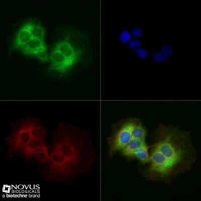
- Immunocytochemistry analysis of DNM1L in HeLa cells fixed for 10 minutes using 10% formalin and then permeabilized for 5 minutes using 1X TBS + 0.5% Triton X-100. Samples were incubated in DNM1L polyclonal antibody (Product # PA1-16987) using a dilution of 5 µg/mL overnight at 4 °C followed by anti-rabbit DyLight 488 (Green) with a dilution of 1:500. Alpha tubulin (DM1A) was used as a co-stain at 1:1000 and detected with an anti-mouse DyLight 550 (Red) at 1:500. Nuclei were counterstained with DAPI (Blue). Cells were imaged using a 40X objective.
- Submitted by
- Invitrogen Antibodies (provider)
- Main image

- Experimental details
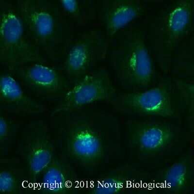
- Immunocytochemistry analysis of DNM1L in PC12 cells fixed for 10 minutes using 10% formalin and then permeabilized for 5 minutes using 1X PBS + 0.05% Triton-X100. Samples were incubated in DNM1L polyclonal antibody (Product # PA1-16987) using a dilution of 2 µg/mL overnight at 4 °C followed by anti-rabbit DyLight 488 (Green) with a dilution of 1:500. Nuclei were counterstained with DAPI (Blue). Cells were imaged using a 40X objective.
Supportive validation
- Submitted by
- Invitrogen Antibodies (provider)
- Main image
- Experimental details
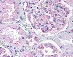
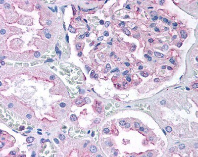
- Immunohistochemical analysis of DNM1L in renal tubular epithelium and visceral epithelial cells of the glomerulus. Samples were incubated in DNM1L polyclonal antibody (Product # PA1-16987). Human kidney cortex, 40X magnification.
- Submitted by
- Invitrogen Antibodies (provider)
- Main image
- Experimental details
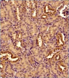
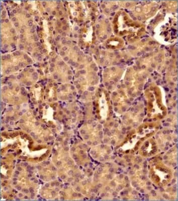
- Immunohistochemical analysis of DNM1L in formalin-fixed paraffin-embedded tissue section of mouse kidney. Samples were incubated in DNM1L polyclonal antibody (Product # PA1-16987) using a dilution of 1:300 followed by a HRP labeled secondary antibody and DAB reagent. Nuclei of the cells were counterstained with hematoxylin. This antibody generated a diffused cytoplasmic staining of DRP1 in the epithelial cells of various tubules and in the cells of glomeruli.
Supportive validation
- Submitted by
- Invitrogen Antibodies (provider)
- Main image
- Experimental details
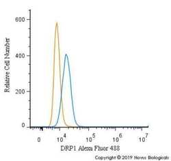
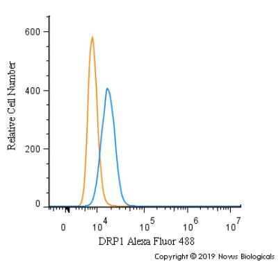
- Flow cytometry of DNM1L in HeLa cells (blue) and a matched isotype control (orange). Samples were incubated in DNM1L polyclonal antibody (Product # PA1-16987) using a dilution of 5 µg/mL for 30 minutes at room temperature. Cells were fixed with 4% PFA and then permeabilized with 0.1% saponin. Both antibodies were conjugated to Alexa Fluor 488.
- Submitted by
- Invitrogen Antibodies (provider)
- Main image
- Experimental details
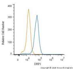
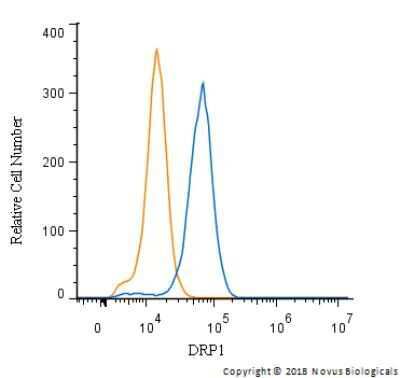
- Flow cytometry of DNM1L in HeLa cells and a matched isotype control. Samples were incubated in DNM1L polyclonal antibody (Product # PA1-16987) using a dilution of 2.5 µg/mL for 30 minutes at room temperature followed by a Rabbit IgG (H+L) Cross-Adsorbed Secondary Antibody, Dylight™ 550. Cells were fixed with 4% PFA and then permeabilized with 0.1% saponin.
Supportive validation
- Submitted by
- Invitrogen Antibodies (provider)
- Main image
- Experimental details
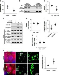
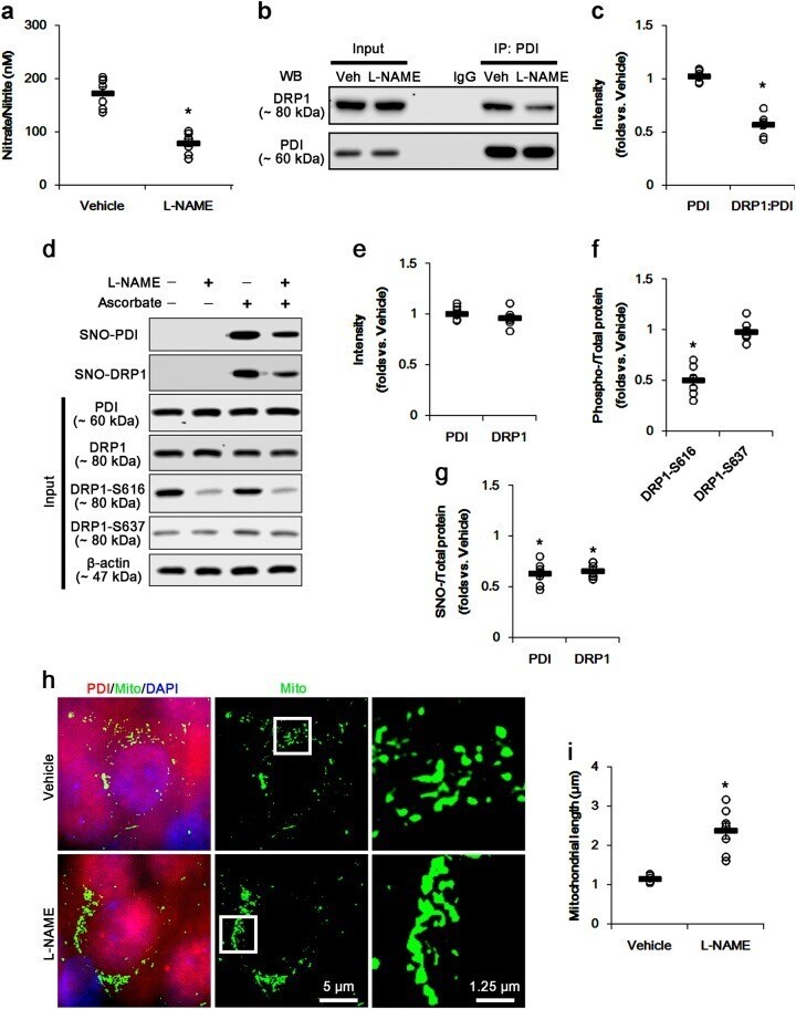
- Fig. 1 The effects of l -NAME on SNO-PDI, SNO-DRP1, and mitochondrial fusion in CA1 neurons under normal condition. l -NAME inhibits NO generation and the binding of PDI to DRP1, and reduces SNO-PDI and SNO-DRP1 levels without changed PDI and DRP1 expressions. However, l -NAME elongates mitochondrial length in CA1 neurons, accompanied by elevated DRP1-S616 phosphorylation. a Quantitative values (mean +- S.E.M) of nitrate/nitrite levels in the hippocampus 7 days after l -NAME infusion. Open circles indicate each individual value. Horizontal bars indicate mean value. Error bars indicate SEM ( *p < 0.05 vs. non-SE animals; n = 7, respectively). b Representative western blot for co-immunoprecipitation of PDI interaction with DRP1 following l -NAME treatment. c Quantification of co-immunoprecipitation analyses of PDI interactions with DRP1 following l -NAME treatment. Open circles indicate each individual value. Horizontal bars indicate mean value. Error bars indicate SEM ( *p < 0.05 vs. vehicle; n = 7, respectively). d Representative western blot for expression, S -nitrosylation and phosphorylation levels of PDI and DRP1 following l -NAME treatment. e - g Quantification of values (mean +- S.E.M) of expression, phosphorylation, and S -nitrosylation levels of PDI and DRP1 following l -NAME treatment. Open circles indicate each individual value. Horizontal bars indicate mean value. Error bars indicate SEM ( *p < 0.05 vs. vehicle; n = 7, respectively). h
- Submitted by
- Invitrogen Antibodies (provider)
- Main image
- Experimental details
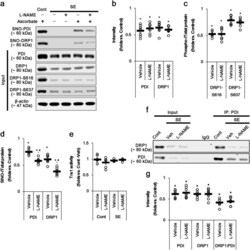
- Fig. 4 The effects of l -NAME on SNO-PDI, SNO-DRP1, Trx1 activity, and mitochondrial fusion in CA1 neurons 3 days after SE. SE significantly reduces expression levels of DRP1 and PDI, PDI-binding to DRP1, and phosphorylation levels of DRP1-S616 and S637 levels independent of Trx1 activity. Both SNO-PDI and SNO-DRP1 are more diminished by l -NAME treatment. a Representative western blot for the effect of l -NAME on expression, S -nitrosylation, and phosphorylation levels of PDI and DRP1 following SE. b - d Quantification of values (mean +- S.E.M) of expression, phosphorylation, and S -nitrosylation levels of PDI and DRP1. Open circles indicate each individual value. Horizontal bars indicate mean value. Error bars indicate SEM (*, # p < 0.05 vs. non-SE animals and vehicle, respectively; n = 7, respectively). e Quantification of values (mean +- S.E.M) of Trx1 activity. Open circles indicate each individual value. Horizontal bars indicate mean value. Error bars indicate SEM ( n = 7, respectively). f Representative western blot for the effect of l -NAME on co-immunoprecipitation of PDI interaction with DRP1 following SE. g Quantification of co-immunoprecipitation analyses of PDI interactions with DRP1. Open circles indicate each individual value. Horizontal bars indicate mean value. Error bars indicate SEM ( *p < 0.05 vs. non-SE animals; n = 7, respectively)

- Submitted by
- Invitrogen Antibodies (provider)
- Main image

- Experimental details
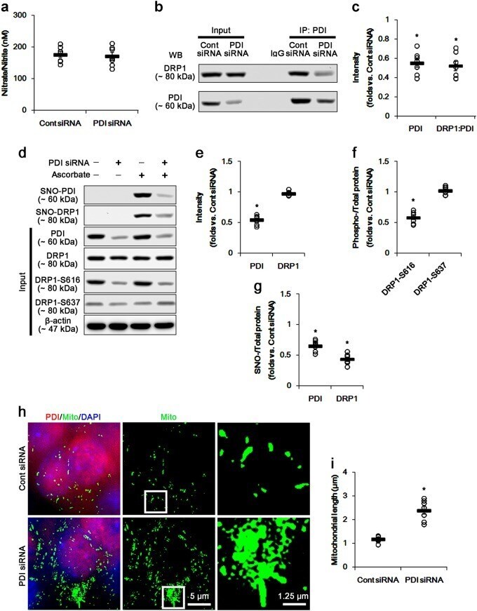
- Fig. 6 The effects of PDI knockdown on SNO-PDI, SNO-DRP1, and mitochondrial fusion in CA1 neurons under normal condition. PDI knockdown reduces PDI expression, the binding of PDI to DRP1, SNO-PDI, and SNO-DRP1 levels without changed DRP1 expression and nitrate/nitrite concentration. However, PDI knockdown elongates mitochondrial length in CA1 neurons, accompanied by elevated DRP1-S616 phosphorylation. a Quantitative values (mean +- S.E.M) of nitrate/nitrite levels in the hippocampus 7 days after PDI siRNA infusion. Open circles indicate each individual value. Horizontal bars indicate mean value. Error bars indicate SEM ( n = 7, respectively). b Representative western blot for co-immunoprecipitation of PDI interaction with DRP1 following PDI knockdown. c Quantification of co-immunoprecipitation analyses of PDI interactions with DRP1 following PDI knockdown. Open circles indicate each individual value. Horizontal bars indicate mean value. Error bars indicate SEM ( *p < 0.05 vs. control siRNA; n = 7, respectively). d Representative western blot for expression, S -nitrosylation, and phosphorylation levels of PDI and DRP1 following PDI knockdown. e - g Quantification of values (mean +- S.E.M) of expression, phosphorylation, and S -nitrosylation levels of PDI and DRP1 following PDI knockdown. Open circles indicate each individual value. Horizontal bars indicate mean value. Error bars indicate SEM ( *p < 0.05 vs. control siRNA; n = 7, respectively). h Quantifica
- Submitted by
- Invitrogen Antibodies (provider)
- Main image

- Experimental details
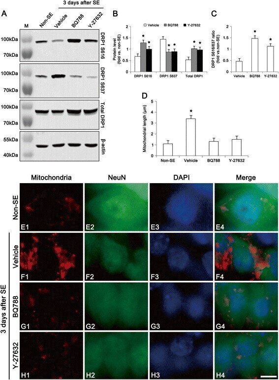
- Fig. 6 Effect of BQ788 and Y-27632 on dysfunction of mitochondrial fission at 3 days after SE. a Western blot images of DRP1, DRP1 S616 and DRP1 S637 in the hippocampus. As compared to vehicle, both BQ788 and Y-27632 (a ROCK inhibitor) attenuate the reductions in DRP1 and DRP1 S616 expression, but increase DRP1 S637 expression. b Quantitative values (mean +- S.E.M) of DRP1, DRP1 S616, DRP1 S637 level ( n = 10 per each group). Significant differences from vehicle, * p < 0.05. c Quantitative values (mean +- S.E.M) of DRP1 S616/S637 ratio in the hippocampus ( n = 10 per each group). Significant differences from vehicle, * p < 0.05. d Quantitative values (mean +- S.E.M) of mitochondrial length in the CA1 neurons ( n = 10 per each group). Significant differences from non-SE animals, * p < 0.05 e-h Representative photographs of mitochondria and NeuN in the CA1 neurons. SE increases mitochondrial length and sphere formation. Both BQ788 and Y-27632 alleviate mitochondrial elongation and sphere formation induced by SE. Bar = 6.25 mum
- Submitted by
- Invitrogen Antibodies (provider)
- Main image
- Experimental details
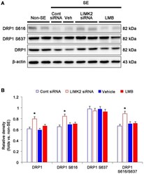
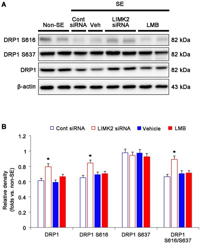
- Figure 4 Effect of LMIK2 siRNA and LMB on HMGB1 expression, DRP1 expression and DRP phosphorylation level at 3 days after SE. (A) Western blot images of HMGB1, DRP1, DRP1 S616 and DRP1 S637 in the hippocampus. Both LIMK2 siRNA and LMB abolish the reduction of HMGB1 release induced by SE. Only LIMK2 siRNA prevents reductions in DRP1 expression, DRP1 S616 level and DRP1 S616/S637 level induced by SE. (B) Quantitative values (mean +- SEM) of HMGB1, DRP1, DRP1 S616, DRP1 S637 level, based on western blot ( n = 7, respectively). * p < 0.05 vs. control siRNA and vehicle, respectively.
- Submitted by
- Invitrogen Antibodies (provider)
- Main image
- Experimental details
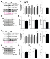
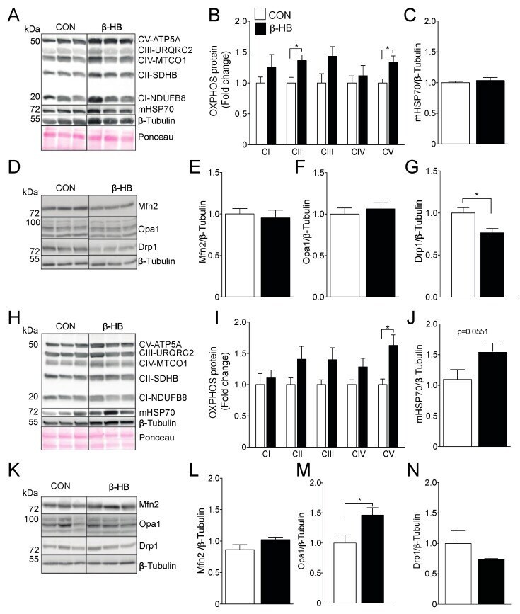
- Figure 2 Six weeks of beta-HB intake alter the electron transport chain and mitochondrial dynamics protein levels. ( A ) Representative OXPHOS Western blot from Tibialis anterior whole tissue extract. ( B ) Tibialis Anterior OXPHOS quantification. ( C ) mHsp70 levels. ( D ) Tibialis anterior mitochondrial fission and fusion proteins representative Western blot. ( E ) Mfn2. ( F ) Opa1 and ( G ) Drp1. ( H ) Soleus representative OXPHOS Western blot. ( I ) Soleus OXPHOS quantification. ( J ) mHsp70. ( K ) Soleus mitochondrial fission and fusion proteins representative Western blot. ( L ) Mfn2. ( M ) Opa1 and ( N ) Drp1. The grouping of blots cropped from different parts of the same gel were divided by black lines. n = 5-6 mice per condition, * p < 0.05, Unpaired t -Test. Values expressed as mean +- SEM.
- Submitted by
- Invitrogen Antibodies (provider)
- Main image
- Experimental details
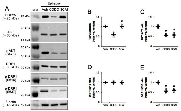
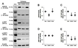
- Effects of CDDO-Me and 3CAI on HSP25, AKT S473, DRP1 S616 and DRP1 S637 levels in the stratum radiatum of the CA1 region of epileptic rats. As compared to vehicle (Veh), CDDO-Me (CDDO) reduces HSP25, AKT S473 and DRP1 S637 levels. 3CAI decreases AKT S473 and DRP1 S637 levels. ( A ) Representative Western blot images of HSP25, AKT, AKT S473, DRP1, DRP1 S616 and DRP1 S637. ( B - E ) Quantifications of HSP25 ( B ), AKT S473 ( C ), DRP1 S616 ( D ) and DRP1 S637 levels. Open circles indicate each value. Horizontal bars indicate the mean value. Error bars indicate SEM (* p < 0.05 vs. Vehicle, respectively; n = 7, respectively).