Explore
 Validate
 Learn
Learn Western blot
 Immunocytochemistry
Antibody data
- Antibody Data
- Antigen structure
- References [2]
- Comments [0]
- Validations
- Immunocytochemistry [2]
- Immunohistochemistry [3]
- Flow cytometry [2]
- Other assay [1]
Submit
Validation data
Reference
Comment
Report error
- Product number
- PA5-79318 - Provider product page

- Provider
- Invitrogen Antibodies
- Product name
- GNAQ Polyclonal Antibody
- Antibody type
- Polyclonal
- Antigen
- Synthetic peptide
- Description
- Reconstitute with 0.2 mL of distilled water to yield a concentration of 500 µg/mL. Positive Control - WB: human placenta tissue, human Jurkat whole cell, rat brain tissue, mouse brain tissue. IHC: mouse testis tissue, rat ovary tissue, human prostatic cancer tissue. ICC/IF: A431 cell. Flow: A431 cell.
- Reactivity
- Human, Mouse, Rat
- Host
- Rabbit
- Isotype
- IgG
- Vial size
- 100 μg
- Concentration
- 500 μg/mL
- Storage
- -20°C
Submitted references Transcriptomic and metabolomic insights into the variety of sperm storage in oviduct of egg layers.
Minute-scale persistence of a GPCR conformation state triggered by non-cognate G protein interactions primes signaling.
Yang G, Li S, Zhao Q, Chu J, Zhou B, Fan S, Shi F, Wei X, Hu X, Zheng X, Liu Z, Zhou X, Tao Y, Li S, Mou C
Poultry science 2021 Jun;100(6):101087
Poultry science 2021 Jun;100(6):101087
Minute-scale persistence of a GPCR conformation state triggered by non-cognate G protein interactions primes signaling.
Gupte TM, Ritt M, Dysthe M, Malik RU, Sivaramakrishnan S
Nature communications 2019 Oct 23;10(1):4836
Nature communications 2019 Oct 23;10(1):4836
No comments: Submit comment
Supportive validation
- Submitted by
- Invitrogen Antibodies (provider)
- Main image
- Experimental details
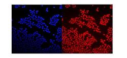
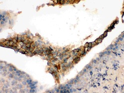
- Immunocytochemistry analysis of GNAQ using anti-GNAQ antibody (Product # PA5-79318). GNAQ was detected in a section of A431 cells. Enzyme antigen retrieval was performed using IHC enzyme antigen retrieval reagent for 15 mins. The cells were blocked with 10% goat serum and then incubated with 2μg/mL rabbit anti-GNAQ antibody (Product # PA5-79318)overnight at 4°C. DyLight®550 Conjugated Goat Anti-Rabbit IgG was used as secondary antibody at 1:100 dilution and incubated for 30 minutes at 37°C. The section was counterstained with DAPI. Visualize using a fluorescence microscope and filter sets appropriate for the label used.

- Submitted by
- Invitrogen Antibodies (provider)
- Main image

- Experimental details
- Immunocytochemistry analysis of GNAQ using anti-GNAQ antibody (Product # PA5-79318). GNAQ was detected in a section of A431 cells. Enzyme antigen retrieval was performed using IHC enzyme antigen retrieval reagent for 15 mins. The cells were blocked with 10% goat serum and then incubated with 2μg/mL rabbit anti-GNAQ antibody (Product # PA5-79318)overnight at 4°C. DyLight®550 Conjugated Goat Anti-Rabbit IgG was used as secondary antibody at 1:100 dilution and incubated for 30 minutes at 37°C. The section was counterstained with DAPI. Visualize using a fluorescence microscope and filter sets appropriate for the label used.

Supportive validation
- Submitted by
- Invitrogen Antibodies (provider)
- Main image
- Experimental details
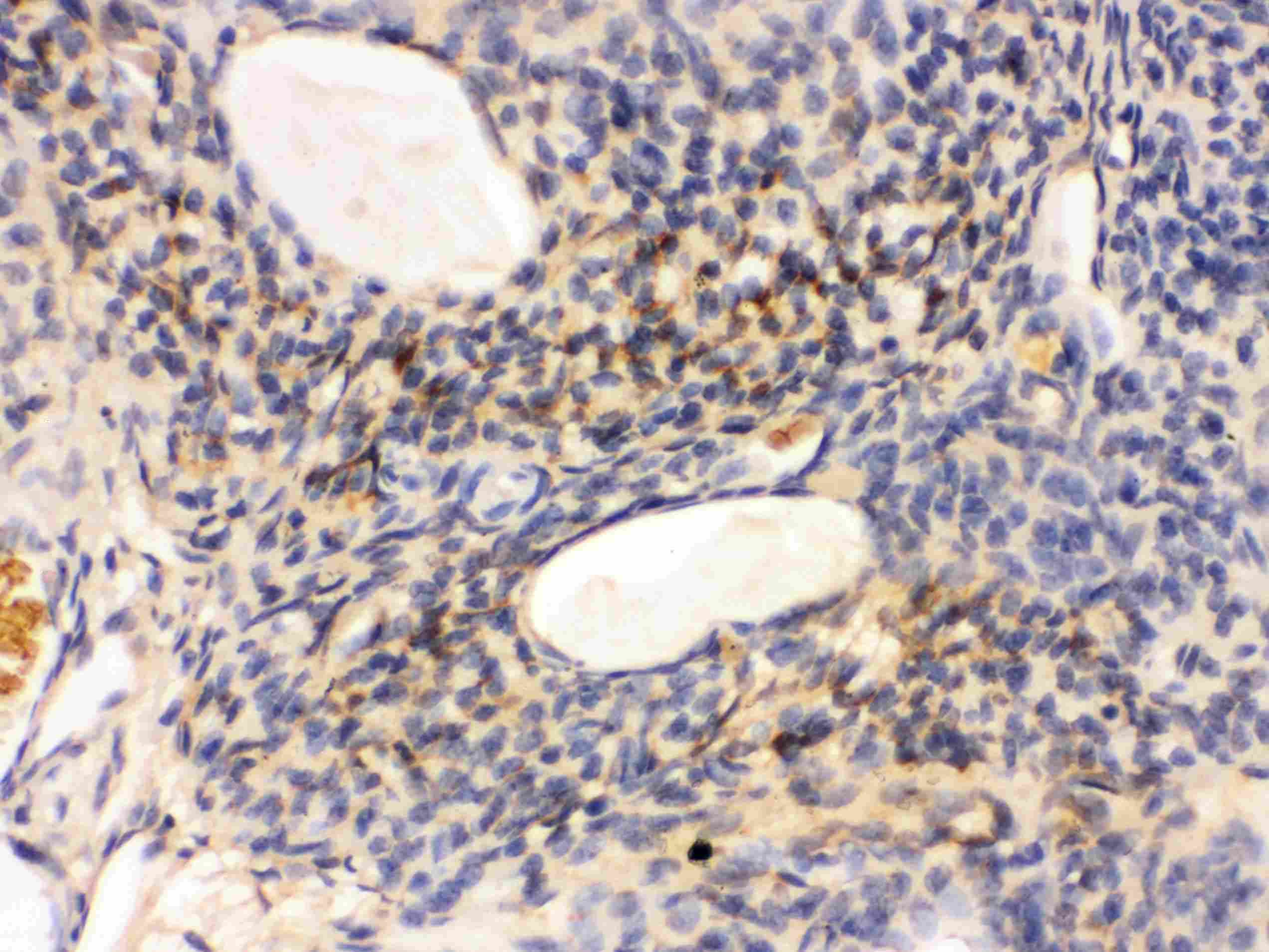
- Immunohistochemistry analysis of GNAQ on paraffin-embedded rat ovary tissue. Sample was incubated with GNAQ polyclonal antibody (Product# PA5-79318).
- Submitted by
- Invitrogen Antibodies (provider)
- Main image

- Experimental details
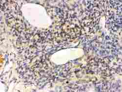
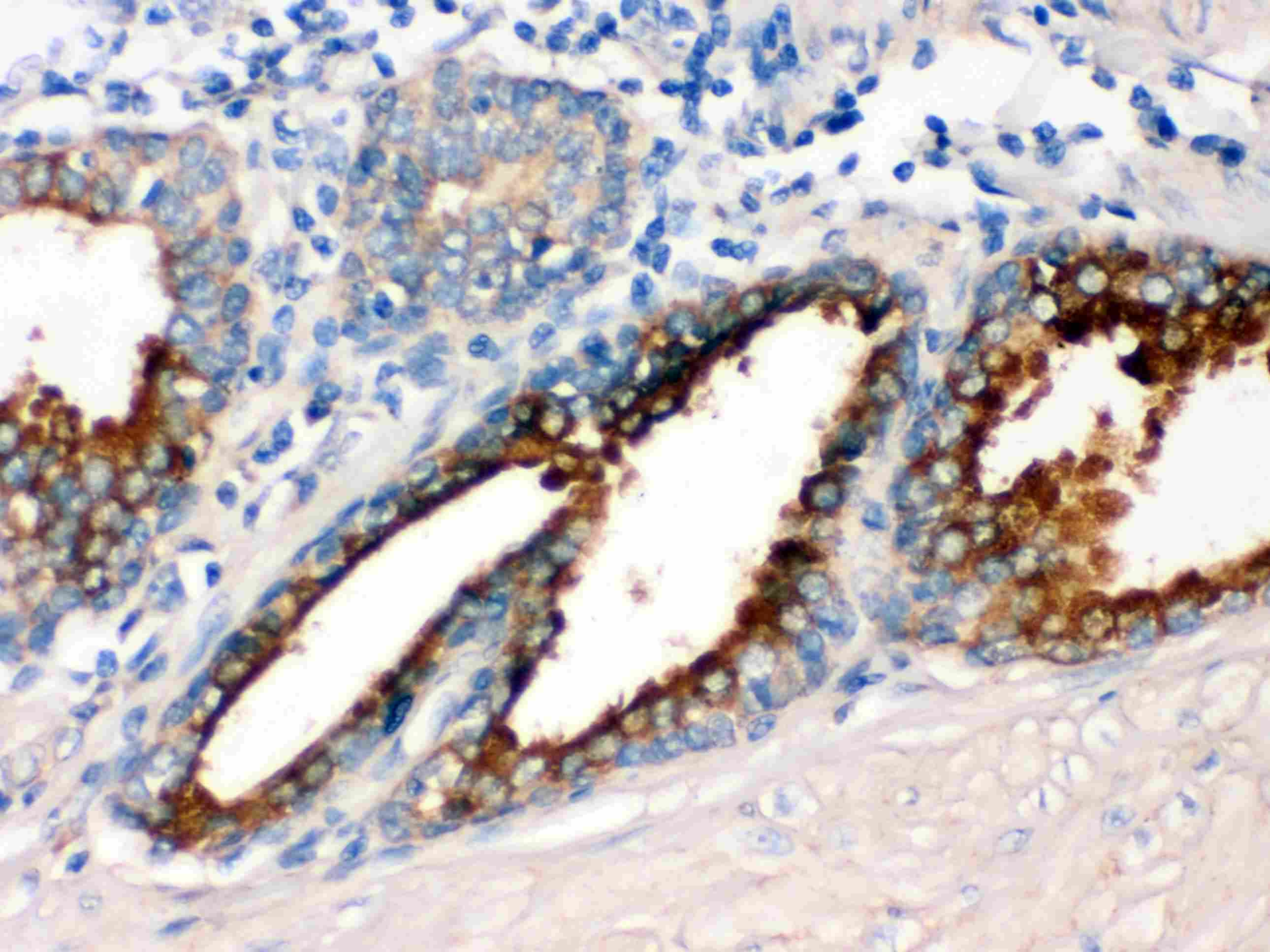
- Immunohistochemistry analysis of GNAQ on paraffin-embedded human prostate cancer tissue. Sample was incubated with GNAQ polyclonal antibody (Product# PA5-79318).
- Submitted by
- Invitrogen Antibodies (provider)
- Main image
- Experimental details
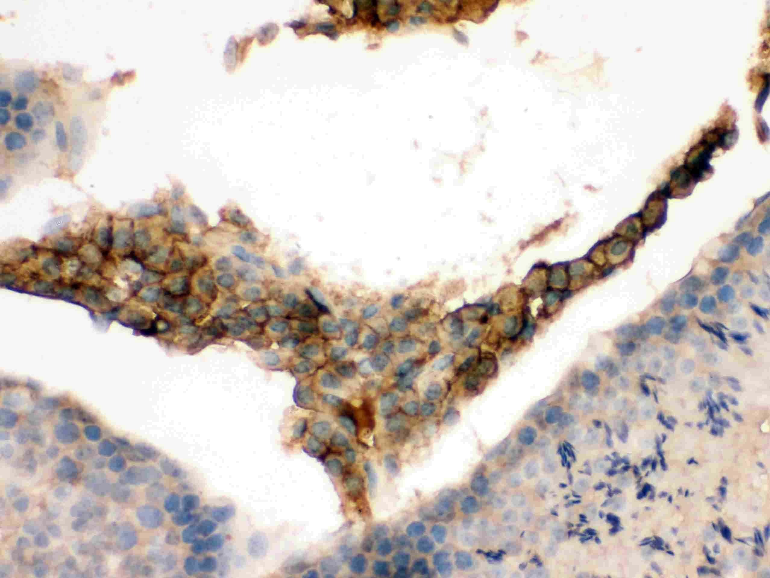
- Immunohistochemistry analysis of GNAQ on paraffin-embedded mouse testis tissue. Sample was incubated with GNAQ polyclonal antibody (Product# PA5-79318).
Supportive validation
- Submitted by
- Invitrogen Antibodies (provider)
- Main image

- Experimental details
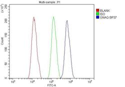
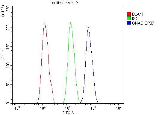
- Flow Cytometry of GNAQ in A431 cells (blue line), isotype control rabbit IgG (green line) and unlabeled (red line). Samples were blocked with 10% goat serum, incubated with GNAQ Polyclonal Antibody (Product # PA5-79318) at a dilution of 1 μg (per 1x10^6 cells), followed by DyLight®488 conjugated goat anti-rabbit IgG (for 30 minutes at 20°C) using 5-10 μg (per 1x10^6 cells) dilution.

- Submitted by
- Invitrogen Antibodies (provider)
- Main image
- Experimental details
- Flow Cytometry of GNAQ in A431 cells (blue line), isotype control rabbit IgG (green line) and unlabeled (red line). Samples were blocked with 10% goat serum, incubated with GNAQ Polyclonal Antibody (Product # PA5-79318) at a dilution of 1 μg (per 1x10^6 cells), followed by DyLight®488 conjugated goat anti-rabbit IgG (for 30 minutes at 20°C) using 5-10 μg (per 1x10^6 cells) dilution.
Supportive validation
- Submitted by
- Invitrogen Antibodies (provider)
- Main image
- Experimental details
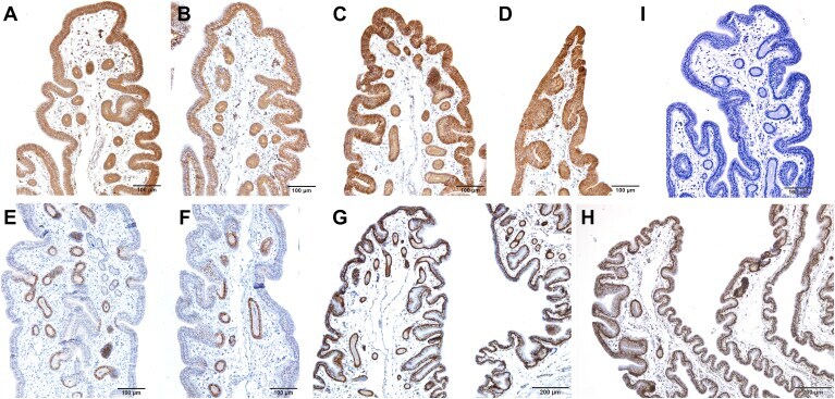
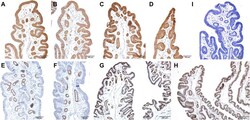
- Figure 4 Immunohistochemistry stained localization of ST6GAL1, GNAQ, ADFP, and PCNA in UVJ of egg layer breeders with high and low sperm storage capacity. (A-B) ST6GAL1 proteins were localized at epithelial cytoplasm in (A) high-SS and (B) low-SS hen UVJ. (C-D) The positive signals of GANQ antibody were localized at epithelial cell membranes in (C) high-SS and (D) low-SS hen UVJ. (E-F) The strong positive signals of ADFP antibody staining were converged on SST in (E) high-SS and (F) low-SS hen UVJ. (G-H) Positive PCNA signals appeared in all epithelial cells in (G) high-SS and (H) low-SS hen UVJ. (I) The negative control without incubating the primary antibody in immunohistochemistry. Abbreviations: SS, sperm storage; SST, sperm storage tubules; UVJ, uterovaginal junction; A-F and I, Bar, 100 mum. G and H, Bar, 200 mum.