Explore
 Validate
 Learn
Learn Western blot
 Immunoprecipitation
Antibody data
- Antibody Data
- Antigen structure
- References [0]
- Comments [0]
- Validations
- Immunoprecipitation [1]
- Immunohistochemistry [2]
- Other assay [1]
Submit
Validation data
Reference
Comment
Report error
- Product number
- MA5-32538 - Provider product page

- Provider
- Invitrogen Antibodies
- Product name
- Midkine Recombinant Rabbit Monoclonal Antibody (JF096-5)
- Antibody type
- Monoclonal
- Antigen
- Synthetic peptide
- Description
- Recombinant rabbit monoclonal antibodies are produced using in vitro expression systems. The expression systems are developed by cloning in the specific antibody DNA sequences from immunoreactive rabbits. Then, individual clones are screened to select the best candidates for production. The advantages of using recombinant rabbit monoclonal antibodies include: better specificity and sensitivity, lot-to-lot consistency, animal origin-free formulations, and broader immunoreactivity to diverse targets due to larger rabbit immune repertoire.
- Reactivity
- Human
- Host
- Rabbit
- Isotype
- IgG
- Antibody clone number
- JF096-5
- Vial size
- 100 μL
- Concentration
- 1 mg/mL
- Storage
- Store at 4°C short term. For long term storage, store at -20°C, avoiding freeze/thaw cycles.
No comments: Submit comment
Supportive validation
- Submitted by
- Invitrogen Antibodies (provider)
- Main image
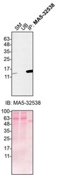
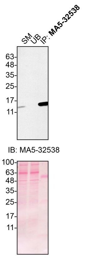
- Experimental details
- HAP1 cells (WT and MDK KO) were washed 3x with PBS and starved for ~18 hrs in DMEM high glucose containing L-glutamate and penicillin/streptomycin. Immunoprecipitation of Midkine was performed by collecting culture media and centrifuging for 10 min at 500 x g to eliminate cells and larger contaminants, then for 10 min at 4500 x g to eliminate smaller contaminants. Culture media were initially concentrated using centrifugal filter units by centrifuging at 4000 x g for 15 min. The resulting 500 µL of the concentrated media were centrifuged again at 4000 x g for 15 min using centrifugal filter units to 200 µL. Antibody-bead conjugates were prepared by adding 1 µg of MDK recombinant monoclonal antibody (Product # MA5-32538) with 30 µL of protein G-Sepharose beads and rocked overnight at 4°C. 0.3 mg of concentrated culture media was incubated with an antibody-bead conjugate for 2 hours at 4°C. Following centrifugation and multiple washes, 10% starting material (SM), 10% unbound fraction (UB) and immunoprecipitated fraction (IP) were processed for immunoblot using the same MDK recombinant monoclonal antibody at 1:200. Ponceau stained transfer of blot is shown. Data courtesy of YCharOS Inc., an open science company with the mission of characterizing commercially available antibodies using knockout validation.

Supportive validation
- Submitted by
- Invitrogen Antibodies (provider)
- Main image
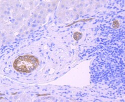
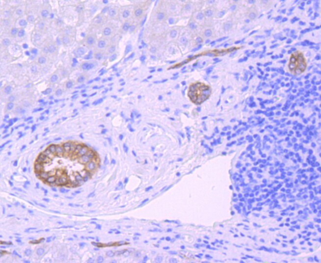
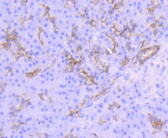
- Experimental details
- Immunohistochemical analysis of Midkine of paraffin-embedded Human liver cancer tissue using a Midkine Monoclonal antibody (Product #MA5-32538). Counter stained with hematoxylin.
- Submitted by
- Invitrogen Antibodies (provider)
- Main image
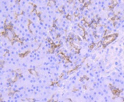
- Experimental details
- Immunohistochemical analysis of Midkine of paraffin-embedded Human pancreas tissue using a Midkine Monoclonal antibody (Product #MA5-32538). Counter stained with hematoxylin.
Supportive validation
- Submitted by
- Invitrogen Antibodies (provider)
- Main image

- Experimental details
- HAP1 cells (WT and MDK KO) were washed 3x with PBS and starved for ~18 hrs in DMEM high glucose containing L-glutamate and penicillin/streptomycin. Immunoprecipitation of Midkine was performed by collecting culture media and centrifuging for 10 min at 500 x g to eliminate cells and larger contaminants, then for 10 min at 4500 x g to eliminate smaller contaminants. Culture media were initially concentrated using centrifugal filter units by centrifuging at 4000 x g for 15 min. The resulting 500 µL of the concentrated media were centrifuged again at 4000 x g for 15 min using centrifugal filter units to 200 µL. Antibody-bead conjugates were prepared by adding 1 µg of MDK recombinant monoclonal antibody (Product # MA5-32538) with 30 µL of protein G-Sepharose beads and rocked overnight at 4°C. 0.3 mg of concentrated culture media was incubated with an antibody-bead conjugate for 2 hours at 4°C. Following centrifugation and multiple washes, 10% starting material (SM), 10% unbound fraction (UB) and immunoprecipitated fraction (IP) were processed for immunoblot using the same MDK recombinant monoclonal antibody at 1:200. Ponceau stained transfer of blot is shown. Data courtesy of YCharOS Inc., an open science company with the mission of characterizing commercially available antibodies using knockout validation.