Explore
 Validate
 Learn
Learn Western blot
Antibody data
- Antibody Data
- Antigen structure
- References [1]
- Comments [0]
- Validations
- Western blot [2]
- Other assay [1]
Submit
Validation data
Reference
Comment
Report error
- Product number
- PA5-30601 - Provider product page

- Provider
- Invitrogen Antibodies
- Product name
- Midkine Polyclonal Antibody
- Antibody type
- Polyclonal
- Antigen
- Recombinant full-length protein
- Description
- Recommended positive controls: NT2D1, U87-MG, MCF-7. Predicted reactivity: Mouse (86%), Rat (86%), Pig (93%), Rabbit (93%), Bovine (92%). Store product as a concentrated solution. Centrifuge briefly prior to opening the vial.
- Reactivity
- Human
- Host
- Rabbit
- Isotype
- IgG
- Vial size
- 100 µL
- Concentration
- 1 mg/mL
- Storage
- Store at 4°C short term. For long term storage, store at -20°C, avoiding freeze/thaw cycles.
Submitted references Expression of ID4 protein in breast cancer cells induces reprogramming of tumour-associated macrophages.
Donzelli S, Milano E, Pruszko M, Sacconi A, Masciarelli S, Iosue I, Melucci E, Gallo E, Terrenato I, Mottolese M, Zylicz M, Zylicz A, Fazi F, Blandino G, Fontemaggi G
Breast cancer research : BCR 2018 Jun 19;20(1):59
Breast cancer research : BCR 2018 Jun 19;20(1):59
No comments: Submit comment
Supportive validation
- Submitted by
- Invitrogen Antibodies (provider)
- Main image
- Experimental details
- Western blot analysis of Midkine using 30 µg of MCF-7 lysate. Samples were loaded onto a 15% SDS-PAGE gel and probed with a Midkine polyclonal antibody (Product # PA5-30601) at a dilution of 1:1000.
- Submitted by
- Invitrogen Antibodies (provider)
- Main image
- Experimental details
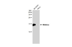
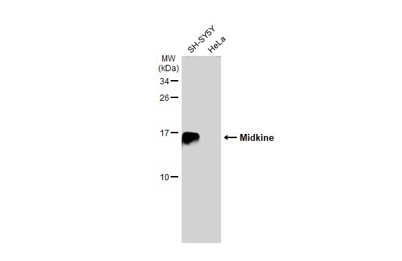
- Western Blot using Midkine Polyclonal Antibody (Product # PA5-30601). Various whole cell extracts (30 µg) were separated by 15% SDS-PAGE, and the membrane was blotted with Midkine Polyclonal Antibody (Product # PA5-30601) diluted at 1:1,000. The HRP-conjugated anti-rabbit IgG antibody was used to detect the primary antibody, and the signal was developed with Trident ECL plus-Enhanced.
Supportive validation
- Submitted by
- Invitrogen Antibodies (provider)
- Main image
- Experimental details
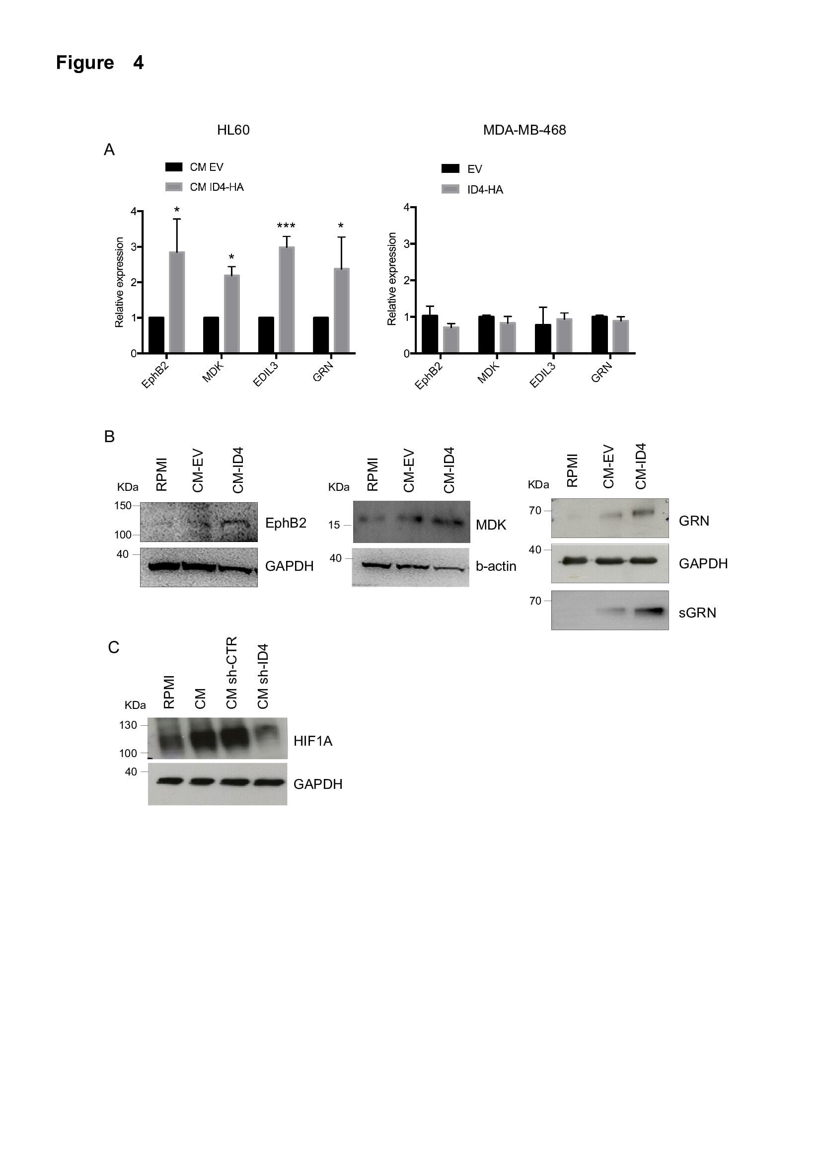
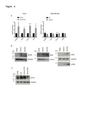
- Additional file 7: Figure S4 a Modulation of selected genes modulated in the TLDA was validated by RT-qPCR in differentiated HL60 cells cultured in CM from ID4-overexpressing (CM ID4-HA) or control (CM EV) MDA-MB-468 cells (left panel). The same transcripts were analysed in MDA-MB-468 cells transfected with ID4-HA expression vector (ID4-HA) or control empty vector (EV) (right panel). b Expression of EphB2, MDK and GRN protein evaluated by Western blotting on lysates from differentiated HL60 cells cultured as in ( a ); secreted GRN (sGRN) was evaluated on CM from differentiated HL60 cells in the same conditions. c HIF1A protein expression evaluated by Western blotting in differentiated U937 cells cultured in RPMI medium or in CM from SKBR3 cells stably interfered for ID4 expression (sh-ID4) or control cells (sh-CTR). (PDF 1320 kb)